Department of Gastroenterology, The First Affiliated Hospital of Nanjing Medical University, Nanjing, 210029, Jiangsu Province, China.
Department of Gastroenterology, The First School of Clinical Medicine of Nanjing Medical University, Nanjing, Jiangsu Province, China.
Dig Dis Sci. 2023 Apr;68(4):1260-1268. doi: 10.1007/s10620-022-07734-y. Epub 2022 Nov 8.
Several studies showed muscularis macrophages (MMφ) are associated with GI motility disorders. The purpose of this study was to preliminary explore the association between MMφ and achalasia.
Tissue samples of the lower esophageal sphincter (LES) high-pressure zone were obtained from 27 achalasia patients and 10 controls. Immunohistochemistry for MMφ, interstitial cells of Cajal (ICC), neuronal nitric oxide synthase (nNOS), and glial cells were conducted. Histological characteristics were compared between groups, and correlation analysis was performed.
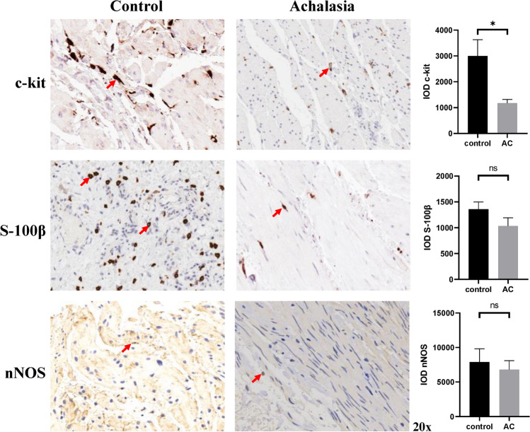
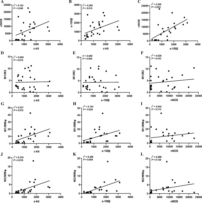
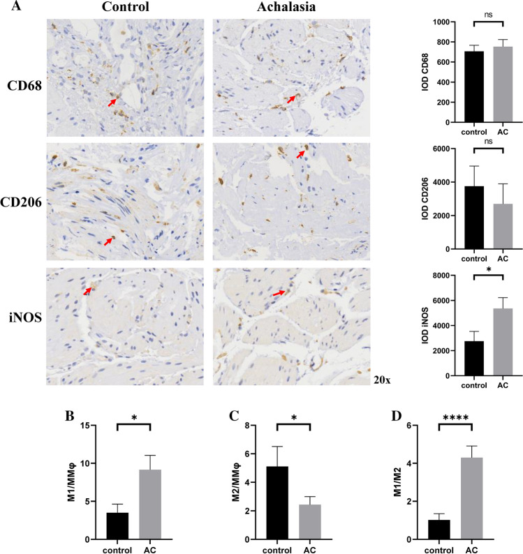
Fewer ICC was found in achalasia compared with controls (P = 0.018), and the level of M1 macrophages was higher than that in controls no matter in terms of the number or the proportion of M1(P = 0.026 for M1 and 0.037 for M1/MMφ). Statistical differences were found between two groups in terms of proportion of M2 and ratio of M1 to M2 (P = 0.048 for M2/ MMφ and < 0.001 for M1/M2). For the correlation analysis, significant correlations were detected between levels of nNOS, ICC, and glial cells in patients with achalasia (P = 0.026 for nNOS and ICC, 0.001 for nNOS and glial cells, 0.019 for ICC and glial cells). There were significant correlations between M2/MMφ and levels of ICC (P = 0.019), glial cells (P = 0.004), and nNOS (P = 0.135).
Patients with achalasia had a higher level of M1/M2 ratio in LES and significant correlations were found between M2/MMφ and numbers of ICC and glial cells, which suggested that MMφ were probably associated with occurrence and development of achalasia.
多项研究表明,肌间巨噬细胞(MMφ)与胃肠道动力障碍有关。本研究旨在初步探讨 MMφ与贲门失弛缓症之间的关系。
从 27 例贲门失弛缓症患者和 10 例对照者的食管下括约肌(LES)高压区获得组织样本。进行 MMφ、Cajal 间质细胞(ICC)、神经元型一氧化氮合酶(nNOS)和神经胶质细胞的免疫组织化学染色。比较两组之间的组织学特征,并进行相关性分析。
与对照组相比,贲门失弛缓症患者的 ICC 数量较少(P = 0.018),且无论从 MMφ 的数量还是 M1 巨噬细胞的比例来看,M1 巨噬细胞的水平均高于对照组(M1 的 P 值分别为 0.026 和 0.037,M1/MMφ 的 P 值为 0.026)。两组之间 M2 的比例和 M1 与 M2 的比值存在统计学差异(M2/MMφ 的 P 值为 0.048,M1/M2 的 P 值 < 0.001)。对于相关性分析,在贲门失弛缓症患者中,nNOS、ICC 和神经胶质细胞的水平之间存在显著相关性(nNOS 与 ICC 的 P 值为 0.026,nNOS 与神经胶质细胞的 P 值为 0.001,ICC 与神经胶质细胞的 P 值为 0.019)。M2/MMφ 与 ICC(P = 0.019)、神经胶质细胞(P = 0.004)和 nNOS(P = 0.135)的水平之间存在显著相关性。
LES 中贲门失弛缓症患者的 M1/M2 比值较高,M2/MMφ 与 ICC 和神经胶质细胞的数量之间存在显著相关性,这提示 MMφ 可能与贲门失弛缓症的发生和发展有关。