Laboratory of Biophysics and Translational Cardiology, Department of Cellular, Computational and Integrative Biology-CIBIO, University of Trento, 38123 Trento, Italy.
CISMed-Centre for Medical Sciences, University of Trento, Trento, Italy.
Europace. 2023 Feb 16;25(2):739-747. doi: 10.1093/europace/euac187.
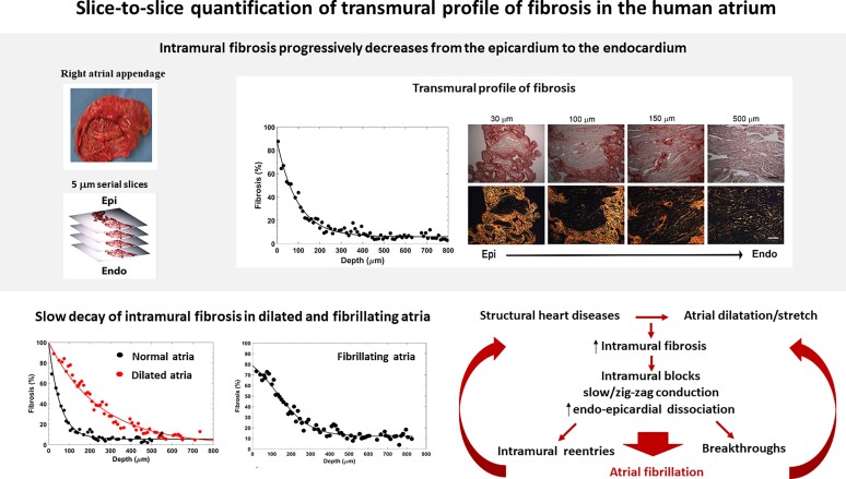
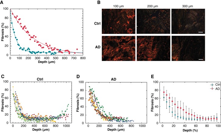
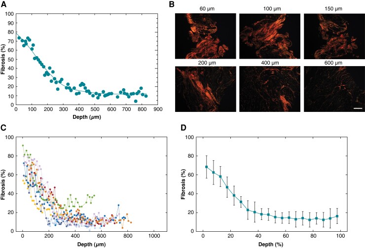
Intramural fibrosis represents a crucial factor in the formation of a three-dimensional (3D) substrate for atrial fibrillation (AF). However, the transmural distribution of fibrosis and its relationship with atrial overload remain largely unknown. The aim of this study is to quantify the transmural profile of atrial fibrosis in patients with different degrees of atrial dilatation and arrhythmic profiles by a high-resolution 3D histology method.
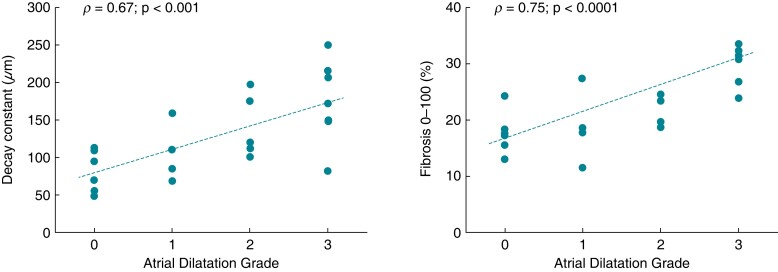
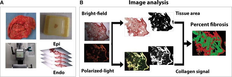
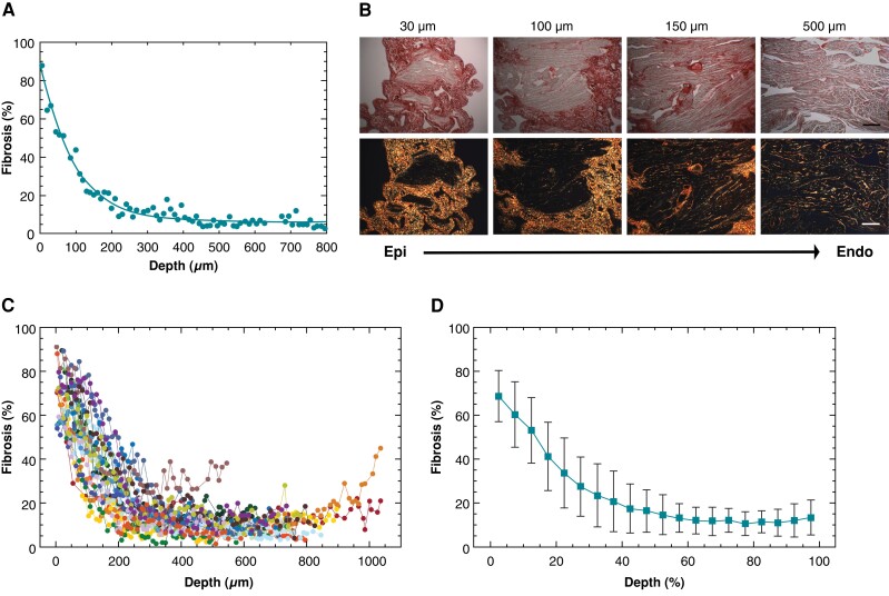
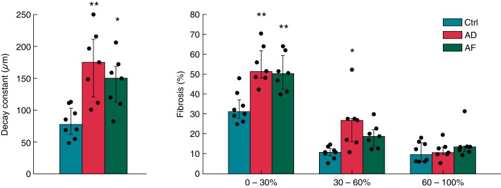
Serial microtome-cut tissue slices, sampling the entire atrial wall thickness at 5 µm spatial resolution, were obtained from right atrial appendage specimens in 23 cardiac surgery patients. Atrial slices were picrosirius red stained, imaged by polarized light microscopy, and analysed by a custom-made segmentation algorithm. In all patients, the intramural fibrosis content displayed a progressive decrease alongside tissue depth, passing from 68.6 ± 11.6% in the subepicardium to 10-13% in the subendocardium. Distinct transmural fibrotic profiles were observed in patients with atrial dilatation with respect to control patients, where the first showed a slower decrease of fibrosis along tissue depth (exponential decay constant: 171.2 ± 54.5 vs. 80.9 ± 24.4 µm, P < 0.005). Similar slow fibrotic profiles were observed in patients with AF (142.8 ± 41.7 µm). Subepicardial and midwall levels of fibrosis correlated with the degree of atrial dilatation (ρ = 0.72, P < 0.001), while no correlation was found in subendocardial layers.
Quantification of fibrosis transmural profile at high resolution is feasible by slice-to-slice histology. Deeper penetration of fibrosis in subepicardial and midwall layers in dilated atria may concur to the formation of a 3D arrhythmic substrate.
壁内纤维化是形成三维(3D)心房颤动(AF)基质的关键因素。然而,纤维化的透壁分布及其与心房负荷的关系在很大程度上尚不清楚。本研究旨在通过高分辨率 3D 组织学方法量化不同程度心房扩张和心律失常患者的心房纤维化透壁分布。
从 23 名心脏手术患者的右心耳标本中获得了连续的超薄切片组织切片,以 5 µm 的空间分辨率采样整个心房壁厚度。心房切片用苦味酸红染色,偏振光显微镜成像,并通过定制的分割算法进行分析。在所有患者中,壁内纤维化含量随组织深度逐渐降低,从心外膜下的 68.6±11.6%降至心内膜下的 10-13%。与对照组患者相比,心房扩张患者表现出不同的透壁纤维化分布,前者沿组织深度的纤维化下降速度较慢(指数衰减常数:171.2±54.5 比 80.9±24.4 µm,P<0.005)。在 AF 患者中也观察到类似的缓慢纤维化分布(142.8±41.7 µm)。心外膜和中层水平的纤维化与心房扩张程度相关(ρ=0.72,P<0.001),而心内膜下各层无相关性。
通过切片到切片的组织学方法,可以对纤维化的透壁分布进行高分辨率的量化。在扩张的心房中,心外膜和中层更深的纤维化渗透可能有助于形成 3D 心律失常基质。