Department of Anatomy, School of Basic Medical Sciences, Anhui Medical University, Hefei, China.
Hefei Cancer Hospital, Chinese Academy of Sciences, Hefei, China.
Invest Ophthalmol Vis Sci. 2022 Nov 1;63(12):16. doi: 10.1167/iovs.63.12.16.
This study aimed to clarify the formation and fixation of the annulus of Zinn (AZ) and its relationship with the extraocular muscles by using ultrathin plastination and three-dimensional models.
Eighteen cadaveric heads (36 sides of the orbital apex) were plastinated to coronal (16 sides), sagittal (13 sides), and horizontal (5 sides) ultrathin plastination sections to be investigated at both macroscopic and microscopic levels. One cadaveric head was used for endoscopic dissection to identify anatomic landmarks.
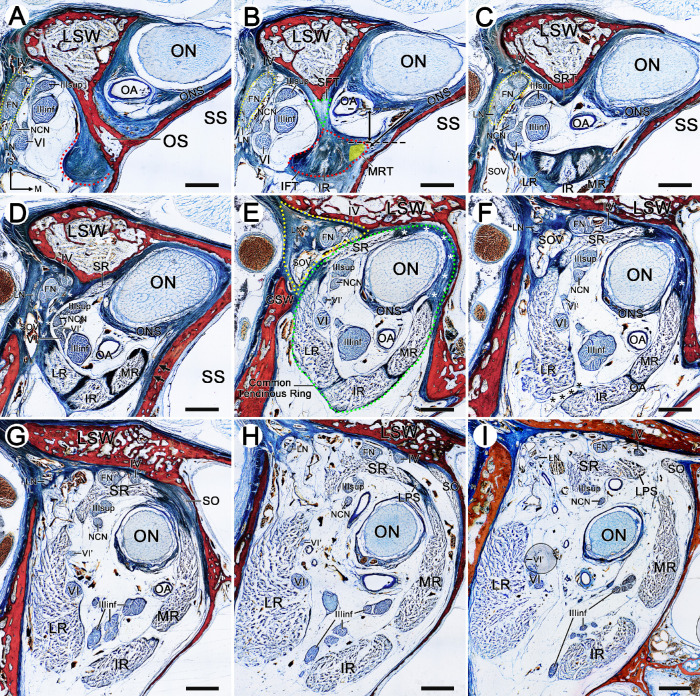
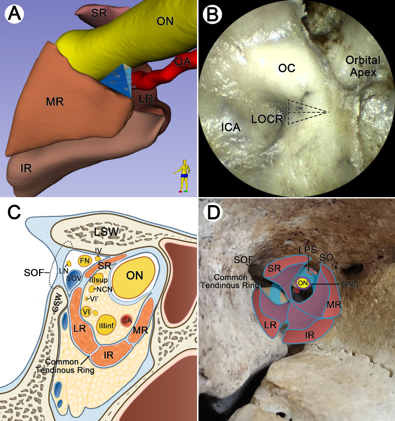
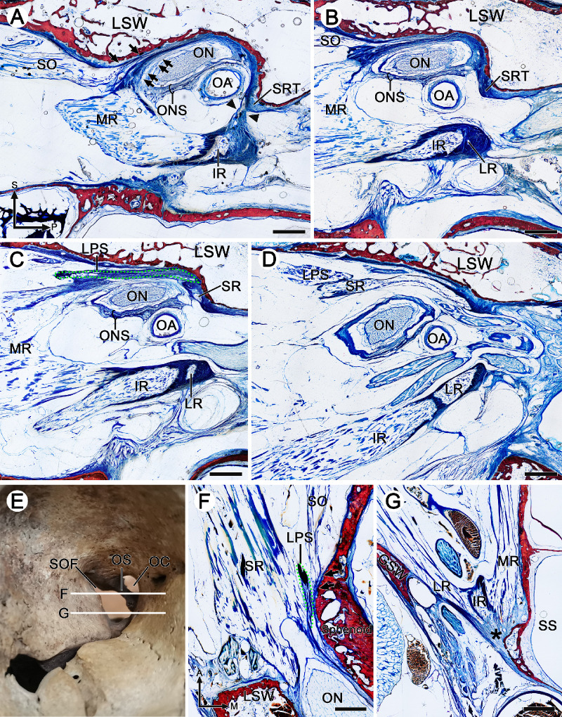
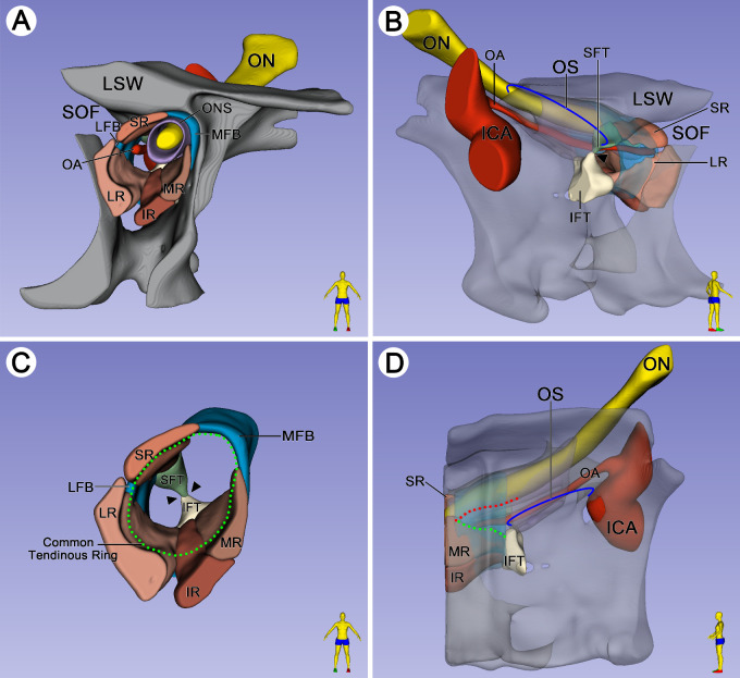
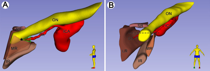
There were two fibrous triangles adhered to both ends of the anterior surface of the optic strut. The superior rectus muscle originated from the superior fibrous triangle, and the lateral, inferior, and medial rectus muscles emerged from the inferior fibrous triangle. It was not until 5.46 ± 0.41 mm anterior to the optic strut that the complete tendinous ring composed of rectus muscles, optic nerve sheath, and periosteum was formed. The superior oblique and levator palpebrae superioris muscles originated from the medial fibrous band of the AZ. At the posterior of the AZ, there was a potential passage between the medial rectus muscle and the optic nerve.
The fixation of the AZ was composed of the connection of the annular tendon to the optic strut posteriorly and the attachment of the complete tendinous ring to the lesser and greater wings of the sphenoid bone anteriorly. The triangular route area between the optic nerve and medial rectus muscle was located on the anterior side of the base of the optic strut.
本研究旨在通过使用超薄塑化和三维模型来阐明 Zinn (AZ)环的形成和固定及其与眼外肌的关系。
18 个头颅(眼眶尖部 36 侧)被塑化成冠状(16 侧)、矢状(13 侧)和水平(5 侧)超薄塑化切片,在宏观和微观水平上进行研究。1 个头颅用于内镜解剖以识别解剖学标志。
在视神经管前表面的两端附着有两个纤维三角。上直肌起源于上纤维三角,而外直肌、下直肌和内直肌则从下纤维三角穿出。直到视神经管前 5.46±0.41mm 处,由直肌、视神经鞘和骨膜组成的完整腱环才形成。上斜肌和上睑提肌起源于 AZ 的内侧纤维带。在 AZ 的后部,内直肌和视神经之间存在潜在的通道。
AZ 的固定由环形腱向后与视神经管的连接以及完整腱环向前附着于蝶骨小翼和大翼组成。视神经和内直肌之间的三角路线区域位于视神经管基部的前侧。