Sevel D
Trans Am Ophthalmol Soc. 1986;84:488-526.
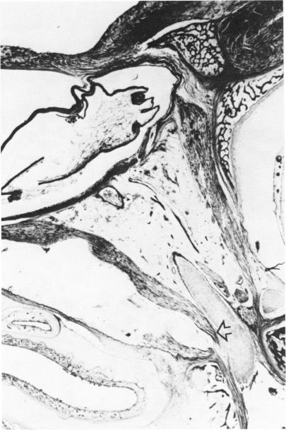
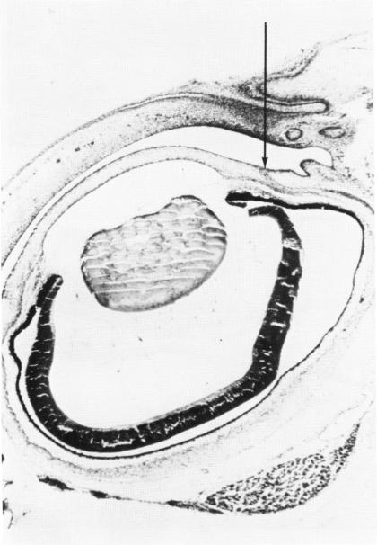
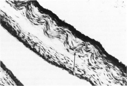
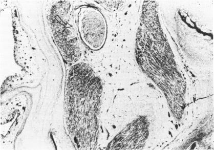
The tendinous origins and insertions of the extraocular muscles were studied embryologically by macroscopic and microscopic methods. It is concluded from this investigation that these tendons of origin and insertion arise from mesenchymal tissue similar to that of their respective muscles. These tendon-muscle groups have developed from superior and inferior mesenchymal complexes. The origins of the extraocular muscles are attached to the periorbita by an interlocking of the tendinous and muscular fibers, which allows for mobility of the extraocular muscles in all extreme directions of gaze and also results in a strong mechanical mooring for these muscles. Avulsion at the origins of the extraocular muscles following severe traction or trauma is rare. The additional origin of the superior and medial rectus muscles to the dura of the optic nerve explains the pain that may occur on movement of the eye in optic neuritis. Optic nerve compression and thyroid myopathy is explained by mucopolysaccharide and inflammatory cell infiltration of the muscular interdigitations that extend up to the site of origin of the rectus muscles. Findings of this investigation suggest that the association of ptosis and superior rectus muscle underaction may be due to a persistence of fibrous tissue that has endured from embryologic development between the superior rectus and levator palpebrae superioris muscles. Superior oblique tendon sheath syndrome is explained by embryologic strands remaining between the tendon of the superior oblique muscle and the trochlea. The insertions of the rectus muscles extend from the equator of the eye to the limbus early on in development. By processes of differential degeneration between the sclera and the rectus tendon, posterior recession of the tendon from the limbus, and contemporaneous growth of the anterior segment of the eye, these tendons reach their adult location only between the ages of 18 months and 2 years. In strabismus surgery, measurements for muscle adjustments should be assessed from the limbus rather than from the sites of insertion of these tendons. In the series of patients with esotropia, no mechanical abnormalities were noted in relationship to the insertions of the medial or lateral recti muscles. Furthermore, no correlation was found between the site of insertion of the medial rectus muscle and the degree of esotropia.
通过宏观和微观方法对眼外肌的腱性起点和止点进行了胚胎学研究。从这项研究得出的结论是,这些起点和止点的肌腱起源于与各自肌肉相似的间充质组织。这些肌腱 - 肌肉群由上、下间充质复合体发育而来。眼外肌的起点通过腱纤维和肌纤维的连锁附着于眶周,这使得眼外肌能够在所有极端注视方向上活动,并且也为这些肌肉提供了强大的机械固定。严重牵引或创伤后眼外肌起点处的撕脱很少见。上直肌和内直肌至视神经硬膜的额外起点解释了视神经炎时眼球运动可能出现的疼痛。视神经受压和甲状腺肌病可通过延伸至直肌起点部位的肌交错处的粘多糖和炎性细胞浸润来解释。这项研究的结果表明,上睑下垂与上直肌作用不足的关联可能是由于上直肌和提上睑肌之间胚胎发育遗留的纤维组织持续存在所致。上斜肌腱鞘综合征可通过上斜肌腱与滑车之间残留的胚胎条索来解释。直肌的止点在发育早期从眼球赤道延伸至角膜缘。通过巩膜和直肌腱之间的差异退变过程、肌腱从角膜缘向后退缩以及眼球前段的同期生长,这些肌腱仅在18个月至2岁之间到达其成人位置。在斜视手术中,肌肉调整的测量应从角膜缘而非这些肌腱的止点部位进行评估。在这组内斜视患者中,未发现与内直肌或外直肌止点相关的机械异常。此外,未发现内直肌止点部位与内斜视程度之间存在相关性。