Biomedical Engineering & Imaging Sciences, King's College London, London SE1 7EH, United Kingdom.
Medical University of Graz, Graz 8036, Austria.
Europace. 2023 Feb 16;25(2):469-477. doi: 10.1093/europace/euac178.
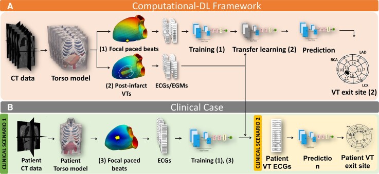
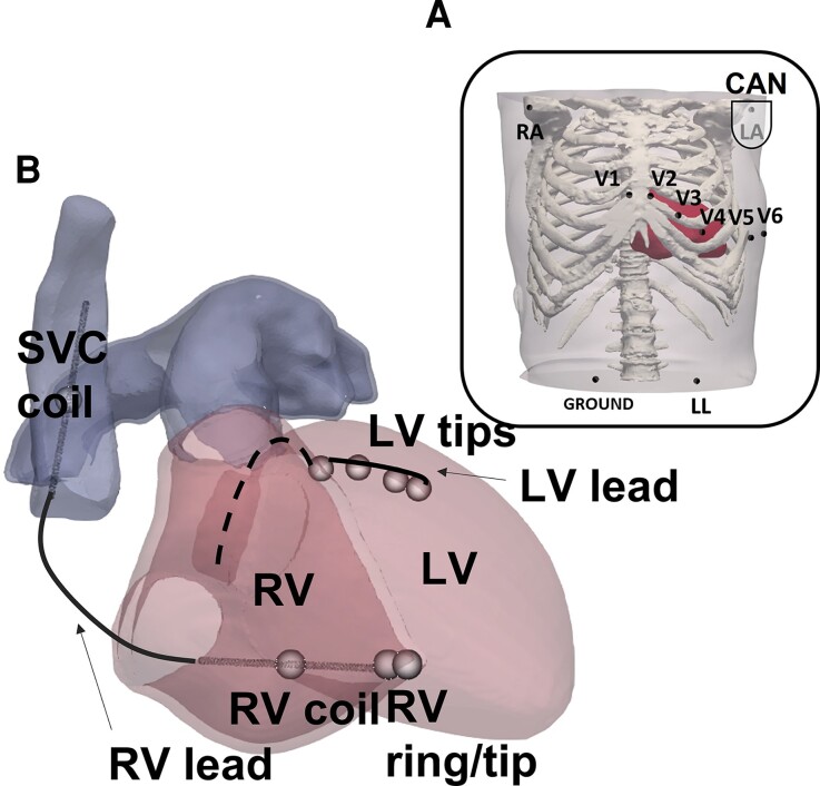
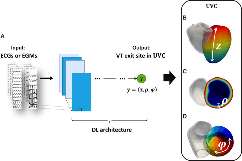
Existing strategies that identify post-infarct ventricular tachycardia (VT) ablation target either employ invasive electrophysiological (EP) mapping or non-invasive modalities utilizing the electrocardiogram (ECG). Their success relies on localizing sites critical to the maintenance of the clinical arrhythmia, not always recorded on the 12-lead ECG. Targeting the clinical VT by utilizing electrograms (EGM) recordings stored in implanted devices may aid ablation planning, enhancing safety and speed and potentially reducing the need of VT induction. In this context, we aim to develop a non-invasive computational-deep learning (DL) platform to localize VT exit sites from surface ECGs and implanted device intracardiac EGMs.
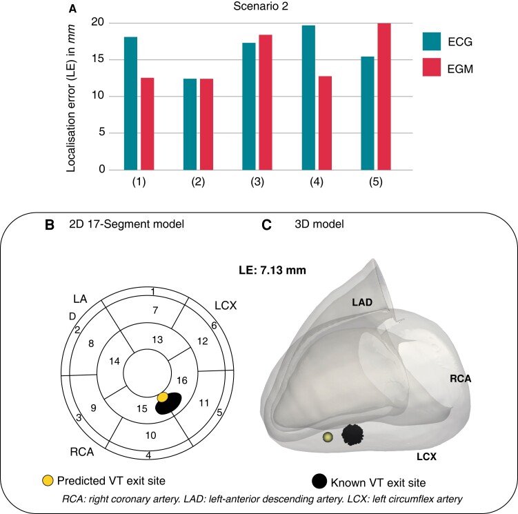
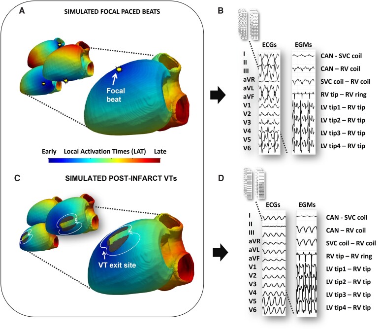
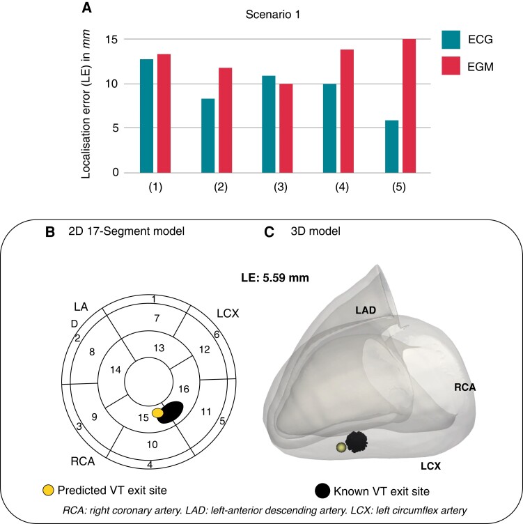
A library of ECGs and EGMs from simulated paced beats and representative post-infarct VTs was generated across five torso models. Traces were used to train DL algorithms to localize VT sites of earliest systolic activation; first tested on simulated data and then on a clinically induced VT to show applicability of our platform in clinical settings. Localization performance was estimated via localization errors (LEs) against known VT exit sites from simulations or clinical ablation targets. Surface ECGs successfully localized post-infarct VTs from simulated data with mean LE = 9.61 ± 2.61 mm across torsos. VT localization was successfully achieved from implanted device intracardiac EGMs with mean LE = 13.10 ± 2.36 mm. Finally, the clinically induced VT localization was in agreement with the clinical ablation volume.
The proposed framework may be utilized for direct localization of post-infarct VTs from surface ECGs and/or implanted device EGMs, or in conjunction with efficient, patient-specific modelling, enhancing safety and speed of ablation planning.
现有的识别心肌梗死后室性心动过速(VT)消融靶点的策略,要么采用侵入性电生理(EP)映射,要么采用利用心电图(ECG)的非侵入性方式。它们的成功依赖于定位对维持临床心律失常至关重要的部位,而这些部位并不总是记录在 12 导联心电图上。通过利用植入设备中存储的心电信号(EGM)记录来针对临床 VT 进行靶向治疗,可能有助于消融计划,提高安全性和速度,并可能减少 VT 诱发的需求。在这种情况下,我们旨在开发一种非侵入性的计算深度学习(DL)平台,以便从体表心电图和植入设备心内 EGM 中定位 VT 出口部位。
在五个躯干模型中生成了模拟起搏搏动和代表性心肌梗死后 VT 的心电图和 EGM 库。这些轨迹用于训练 DL 算法以定位 VT 最早收缩激活部位;首先在模拟数据上进行测试,然后在临床诱导的 VT 上进行测试,以展示我们平台在临床环境中的适用性。通过与模拟中的 VT 出口部位或临床消融靶点的定位误差(LE)来估计定位性能。体表心电图成功地从模拟数据中定位了心肌梗死后 VT,跨躯干的平均 LE 为 9.61±2.61mm。从植入设备心内 EGM 成功地定位了 VT,平均 LE 为 13.10±2.36mm。最后,临床诱导的 VT 定位与临床消融体积一致。
所提出的框架可用于直接从体表心电图和/或植入设备 EGM 定位心肌梗死后 VT,或与高效、患者特异性建模结合使用,增强消融计划的安全性和速度。