Mendel Centre for Genomics and Proteomics of Plants, Central European Institute of Technology (CEITEC), Masaryk University, Brno, Czech Republic.
National Centre for Biomolecular Research, Faculty of Science, Masaryk University, Brno, Czech Republic.
Plant Reprod. 2022 Dec;35(4):279-293. doi: 10.1007/s00497-022-00453-4. Epub 2022 Nov 15.
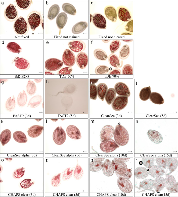
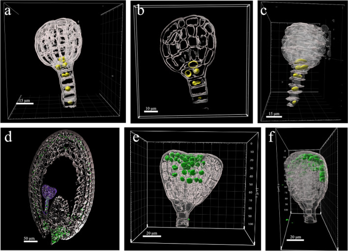
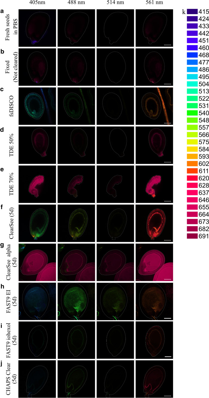
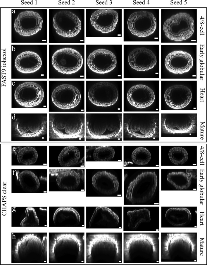
ClearSee alpha and FAST9 were optimized for imaging Arabidopsis seeds up to the torpedo stages. The methods preserve the fluorescence of reporter proteins and seed shape, allowing phenotyping embryos in intact seeds. Tissue clearing methods eliminate the need for sectioning, thereby helping better understand the 3D organization of tissues and organs. In the past fifteen years, clearing methods have been developed to preserve endogenous fluorescent protein tags. Some of these methods (ClearSee, TDE, PEA-Clarity, etc.) were adapted to clear various plant species, with the focus on roots, leaves, shoot apical meristems, and floral parts. However, these methods have not been used in developing seeds beyond the early globular stage. Tissue clearing is problematic in post-globular seeds due to various apoplastic barriers and secondary metabolites. In this study, we compared six methods for their efficiency in clearing Arabidopsis thaliana seeds at post-globular embryonic stages. Three methods (TDE, ClearSee, and ClearSee alpha) have already been reported in plants, whereas the others (fsDISCO, FAST9, and CHAPS clear) are used in this context for the first time. These methods were assessed for seed morphological changes, clearing capacity, removal of tannins, and spectral properties. We tested each method in seeds from globular to mature stages. The pros and cons of each method are listed herein. ClearSee alpha appears to be the method of choice as it preserves seed morphology and prevents tannin oxidation. However, FAST9 with 60% iohexol as a mounting medium is faster, clears better, and appears suitable for embryonic shape imaging. Our results may guide plant researchers to choose a suitable method for imaging fluorescent protein-labeled embryos in intact Arabidopsis seeds.
ClearSee alpha 和 FAST9 经过优化,可用于对拟南芥种子进行成像,直至鱼雷阶段。这些方法保留了报告蛋白和种子形状的荧光,使我们能够对完整种子中的胚胎进行表型分析。组织透明化方法无需进行切片,从而有助于更好地理解组织和器官的 3D 结构。在过去的十五年中,已经开发了多种方法来保留内源性荧光蛋白标签。其中一些方法(ClearSee、TDE、PEA-Clarity 等)已被用于透明化各种植物物种,重点是根、叶、茎尖分生组织和花器官。然而,这些方法在种子发育到早期球形阶段之后并未被用于开发种子。由于存在各种质外体屏障和次生代谢物,在球形后期的种子中进行组织透明化存在问题。在本研究中,我们比较了六种方法在透明化拟南芥种子后期球形胚胎阶段的效率。其中三种方法(TDE、ClearSee 和 ClearSee alpha)已经在植物中报道过,而另外三种方法(fsDISCO、FAST9 和 CHAPS clear)则是首次在这种情况下使用。我们评估了这些方法对种子形态变化、透明化能力、单宁去除和光谱特性的影响。我们在从球形期到成熟阶段的种子中测试了每种方法。在此列出了每种方法的优缺点。ClearSee alpha 似乎是首选方法,因为它可以保持种子形态并防止单宁氧化。然而,使用 60% iohexol 作为载剂的 FAST9 速度更快,透明化效果更好,似乎适合用于胚胎形状成像。我们的结果可能为植物研究人员选择合适的方法来对完整的拟南芥种子中的荧光蛋白标记的胚胎进行成像提供指导。