• 文献检索
  • 文档翻译
  • 深度研究
  • 学术资讯
  • Suppr Zotero 插件Zotero 插件
  • 邀请有礼
  • 套餐&价格
  • 历史记录
应用&插件
Suppr Zotero 插件Zotero 插件浏览器插件Mac 客户端Windows 客户端微信小程序
定价
高级版会员购买积分包购买API积分包
服务
文献检索文档翻译深度研究API 文档MCP 服务
关于我们
关于 Suppr公司介绍联系我们用户协议隐私条款
关注我们

Suppr 超能文献

核心技术专利:CN118964589B侵权必究
粤ICP备2023148730 号-1Suppr @ 2026

文献检索

告别复杂PubMed语法,用中文像聊天一样搜索,搜遍4000万医学文献。AI智能推荐,让科研检索更轻松。

立即免费搜索

文件翻译

保留排版,准确专业,支持PDF/Word/PPT等文件格式,支持 12+语言互译。

免费翻译文档

深度研究

AI帮你快速写综述,25分钟生成高质量综述,智能提取关键信息,辅助科研写作。

立即免费体验

基底动脉粥样硬化斑块的高危特征。

High-risk features of basilar artery atherosclerotic plaque.

作者信息

Li Shaojun, Wei Jiana, Huang Ruiyun, Li Chenghao, Chen Hongbing, Qiu Zhihua, Jiang Yongjun, Wu Li

机构信息

Department of Neurology, The Second Affiliated Hospital of Guangzhou Medical University, Guangzhou, China.

Department of Neurology and Stroke Center, The First Affiliated Hospital, Sun Yat-sen University, Guangzhou, China.

出版信息

Front Neurol. 2022 Oct 28;13:1019036. doi: 10.3389/fneur.2022.1019036. eCollection 2022.

DOI:10.3389/fneur.2022.1019036
PMID:36388175
原文链接:https://pmc.ncbi.nlm.nih.gov/articles/PMC9650051/
Abstract

INTRODUCTION

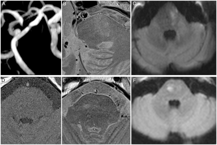
High-resolution magnetic resonance imaging (HR-MRI) is used to characterize atherosclerotic plaque. The present study aimed to determine the high-risk features of the basilar artery (BA) atherosclerotic plaque.

METHODS

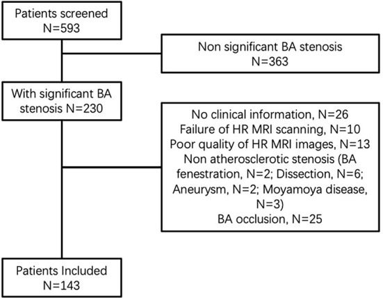
Patients with advanced BA stenosis were screened. The features including the ruptured fibrous cap (RFC), lipid core, intraplaque hemorrhage (IPH), plaque enhancement, and calcification were assessed by using high-resolution MRI. The relationship between the features and acute infarction was analyzed.

RESULTS

From 1 June 2014 to 31 December 2018, a total of 143 patients with 76 new strokes were included. RFC was identified in 25% of symptomatic and 10.4% of asymptomatic patients. IPH was identified in 48.7% of symptomatic and 25.4% of asymptomatic patients. RFC (3.157, 95% CI 1.062 to 9.382, = 0.039) and IPH (2.78, 95% CI 1.127 to 6.505, = 0.026) were independent risk factors for acute infarction.

CONCLUSION

Our study showed that RFC and IPH of BA plaque were independent risk factors for acute infarction.

摘要

引言

高分辨率磁共振成像(HR-MRI)用于表征动脉粥样硬化斑块。本研究旨在确定基底动脉(BA)粥样硬化斑块的高危特征。

方法

筛选患有晚期BA狭窄的患者。使用高分辨率MRI评估包括纤维帽破裂(RFC)、脂质核心、斑块内出血(IPH)、斑块强化和钙化等特征。分析这些特征与急性梗死之间的关系。

结果

2014年6月1日至2018年12月31日,共纳入143例患者,其中76例发生新的卒中。有症状患者中25%发现RFC,无症状患者中10.4%发现RFC。有症状患者中48.7%发现IPH,无症状患者中25.4%发现IPH。RFC(3.157,95%可信区间1.062至9.382,P = 0.039)和IPH(2.78,95%可信区间1.127至6.505,P = 0.026)是急性梗死的独立危险因素。

结论

我们的研究表明,BA斑块的RFC和IPH是急性梗死的独立危险因素。

https://cdn.ncbi.nlm.nih.gov/pmc/blobs/9355/9650051/ff84d1a72df2/fneur-13-1019036-g0002.jpg
https://cdn.ncbi.nlm.nih.gov/pmc/blobs/9355/9650051/4d4f370aa566/fneur-13-1019036-g0001.jpg
https://cdn.ncbi.nlm.nih.gov/pmc/blobs/9355/9650051/ff84d1a72df2/fneur-13-1019036-g0002.jpg
https://cdn.ncbi.nlm.nih.gov/pmc/blobs/9355/9650051/4d4f370aa566/fneur-13-1019036-g0001.jpg
https://cdn.ncbi.nlm.nih.gov/pmc/blobs/9355/9650051/ff84d1a72df2/fneur-13-1019036-g0002.jpg

相似文献

1
High-risk features of basilar artery atherosclerotic plaque.基底动脉粥样硬化斑块的高危特征。
Front Neurol. 2022 Oct 28;13:1019036. doi: 10.3389/fneur.2022.1019036. eCollection 2022.
2
MR imaging of vulnerable carotid plaque.易损性颈动脉斑块的磁共振成像
Cardiovasc Diagn Ther. 2020 Aug;10(4):1019-1031. doi: 10.21037/cdt.2020.03.12.
3
Contemporary carotid imaging: from degree of stenosis to plaque vulnerability.当代颈动脉成像:从狭窄程度到斑块易损性
J Neurosurg. 2016 Jan;124(1):27-42. doi: 10.3171/2015.1.JNS142452. Epub 2015 Jul 31.
4
Association of Intraplaque Hemorrhage and Acute Infarction in Patients With Basilar Artery Plaque.基底动脉斑块患者斑块内出血与急性梗死的相关性
Stroke. 2015 Oct;46(10):2768-72. doi: 10.1161/STROKEAHA.115.009412. Epub 2015 Aug 25.
5
Identification of high-risk plaque features in intracranial atherosclerosis: initial experience using a radiomic approach.颅内动脉粥样硬化斑块高危特征的识别:一种基于放射组学方法的初步经验。
Eur Radiol. 2018 Sep;28(9):3912-3921. doi: 10.1007/s00330-018-5395-1. Epub 2018 Apr 9.
6
Characteristics of Symptomatic Basilar Artery Stenosis Using High-Resolution Magnetic Resonance Imaging in Ischemic Stroke Patients.症状性基底动脉狭窄的特征:基于缺血性脑卒中患者的高分辨率磁共振成像研究。
J Atheroscler Thromb. 2021 Oct 1;28(10):1063-1070. doi: 10.5551/jat.58214. Epub 2020 Nov 15.
7
Intraplaque hemorrhage, fibrous cap status, and microembolic signals in symptomatic patients with mild to moderate carotid artery stenosis: the Plaque at RISK study.症状性轻至中度颈动脉狭窄患者的斑块内出血、纤维帽状态及微栓塞信号:高危斑块研究
Stroke. 2014 Nov;45(11):3423-6. doi: 10.1161/STROKEAHA.114.006800. Epub 2014 Sep 25.
8
Clinical Significance of Intraplaque Hemorrhage in Low- and High-Grade Basilar Artery Stenosis on High-Resolution MRI.高分辨率 MRI 显示低级别和高级别基底动脉狭窄斑块内出血的临床意义。
AJNR Am J Neuroradiol. 2018 Jul;39(7):1286-1292. doi: 10.3174/ajnr.A5676. Epub 2018 May 24.
9
Sex Differences in Plaque Composition and Morphology Among Symptomatic Patients With Mild-to-Moderate Carotid Artery Stenosis.症状性轻至中度颈动脉狭窄患者斑块成分和形态的性别差异。
Stroke. 2022 Feb;53(2):370-378. doi: 10.1161/STROKEAHA.121.036564. Epub 2022 Jan 5.
10
Normalized wall index, intraplaque hemorrhage and ulceration of carotid plaques correlate with the severity of ischemic stroke.颈动脉斑块的管壁归一化指数、斑块内出血和溃疡与缺血性脑卒中的严重程度相关。
Atherosclerosis. 2020 Dec;315:138-144. doi: 10.1016/j.atherosclerosis.2020.10.896. Epub 2020 Nov 6.

引用本文的文献

1
Characteristics and evaluation of atherosclerotic plaques: an overview of state-of-the-art techniques.动脉粥样硬化斑块的特征与评估:最新技术综述
Front Neurol. 2023 Oct 12;14:1159288. doi: 10.3389/fneur.2023.1159288. eCollection 2023.
2
The Role of High-Resolution Magnetic Resonance Imaging in Cerebrovascular Disease: A Narrative Review.高分辨率磁共振成像在脑血管疾病中的作用:一篇叙述性综述。
Brain Sci. 2023 Apr 18;13(4):677. doi: 10.3390/brainsci13040677.

本文引用的文献

1
High-resolution MRI of intracranial large artery diseases: how to use it in clinical practice?颅内大动脉疾病的高分辨率 MRI:如何在临床实践中应用?
Stroke Vasc Neurol. 2019 Jun 20;4(2):102-104. doi: 10.1136/svn-2018-000210. eCollection 2019 Jul.
2
Lipid-Rich Necrotic Core of Basilar Artery Atherosclerotic Plaque: Contrast-Enhanced Black Blood Imaging on Vessel Wall Imaging.基底动脉粥样硬化斑块富含脂质的坏死核心:血管壁成像上的对比增强黑血成像
Diagnostics (Basel). 2019 Jul 2;9(3):69. doi: 10.3390/diagnostics9030069.
3
Size of carotid artery intraplaque hemorrhage and acute ischemic stroke: a cardiovascular magnetic resonance Chinese atherosclerosis risk evaluation study.
颈动脉斑块内出血与急性缺血性脑卒中的大小:一项心血管磁共振中国动脉粥样硬化风险评估研究。
J Cardiovasc Magn Reson. 2019 Jul 1;21(1):36. doi: 10.1186/s12968-019-0548-1.
4
Intravascular Optical Coherence Tomography for Neurointerventional Surgery.用于神经介入手术的血管内光学相干断层扫描
Stroke. 2019 Jan;50(1):218-223. doi: 10.1161/STROKEAHA.118.022315. Epub 2018 Nov 29.
5
Relationship between the geometry patterns of vertebrobasilar artery and atherosclerosis.椎基底动脉几何形态与动脉粥样硬化之间的关系。
BMC Neurol. 2018 Jun 12;18(1):83. doi: 10.1186/s12883-018-1084-6.
6
Clinical Significance of Intraplaque Hemorrhage in Low- and High-Grade Basilar Artery Stenosis on High-Resolution MRI.高分辨率 MRI 显示低级别和高级别基底动脉狭窄斑块内出血的临床意义。
AJNR Am J Neuroradiol. 2018 Jul;39(7):1286-1292. doi: 10.3174/ajnr.A5676. Epub 2018 May 24.
7
Posterior circulation ischaemic stroke.后循环缺血性卒中
BMJ. 2018 Apr 19;361:k1185. doi: 10.1136/bmj.k1185.
8
Identification of high-risk plaque features in intracranial atherosclerosis: initial experience using a radiomic approach.颅内动脉粥样硬化斑块高危特征的识别:一种基于放射组学方法的初步经验。
Eur Radiol. 2018 Sep;28(9):3912-3921. doi: 10.1007/s00330-018-5395-1. Epub 2018 Apr 9.
9
Incremental Value of Plaque Enhancement in Patients with Moderate or Severe Basilar Artery Stenosis: 3.0 T High-Resolution Magnetic Resonance Study.中重度基底动脉狭窄患者斑块强化的增量价值:3.0T 高分辨率磁共振研究。
Biomed Res Int. 2017;2017:4281629. doi: 10.1155/2017/4281629. Epub 2017 Sep 17.
10
Carotid Plaque Lipid Content and Fibrous Cap Status Predict Systemic CV Outcomes: The MRI Substudy in AIM-HIGH.颈动脉斑块脂质含量和纤维帽状态预测全身性心血管结局:AIM-HIGH研究中的MRI子研究
JACC Cardiovasc Imaging. 2017 Mar;10(3):241-249. doi: 10.1016/j.jcmg.2016.06.017.