• 文献检索
  • 文档翻译
  • 深度研究
  • 学术资讯
  • Suppr Zotero 插件Zotero 插件
  • 邀请有礼
  • 套餐&价格
  • 历史记录
应用&插件
Suppr Zotero 插件Zotero 插件浏览器插件Mac 客户端Windows 客户端微信小程序
定价
高级版会员购买积分包购买API积分包
服务
文献检索文档翻译深度研究API 文档MCP 服务
关于我们
关于 Suppr公司介绍联系我们用户协议隐私条款
关注我们

Suppr 超能文献

核心技术专利:CN118964589B侵权必究
粤ICP备2023148730 号-1Suppr @ 2026

文献检索

告别复杂PubMed语法,用中文像聊天一样搜索,搜遍4000万医学文献。AI智能推荐,让科研检索更轻松。

立即免费搜索

文件翻译

保留排版,准确专业,支持PDF/Word/PPT等文件格式,支持 12+语言互译。

免费翻译文档

深度研究

AI帮你快速写综述,25分钟生成高质量综述,智能提取关键信息,辅助科研写作。

立即免费体验

两种荧光探针染色方法对生物膜细胞活力评估及可视化的比较

Comparison of cell viability assessment and visualization of biofilm with two fluorescent probe staining methods.

作者信息

Shailaja Aswathy, Bruce Terri F, Gerard Patrick, Powell Rhonda R, Pettigrew Charles A, Kerrigan Julia L

机构信息

Department of Plant and Environmental Sciences, Clemson University, Clemson, SC, USA.

Clemson Light Imaging Facility, Clemson University, Clemson, SC, USA.

出版信息

Biofilm. 2022 Oct 28;4:100090. doi: 10.1016/j.bioflm.2022.100090. eCollection 2022 Dec.

DOI:10.1016/j.bioflm.2022.100090
PMID:36389263
原文链接:https://pmc.ncbi.nlm.nih.gov/articles/PMC9646680/
Abstract

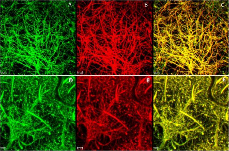
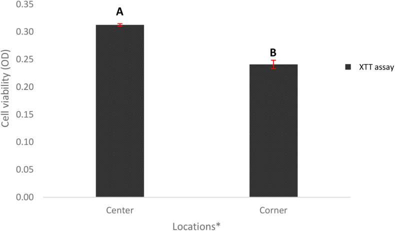
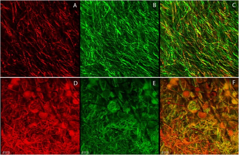
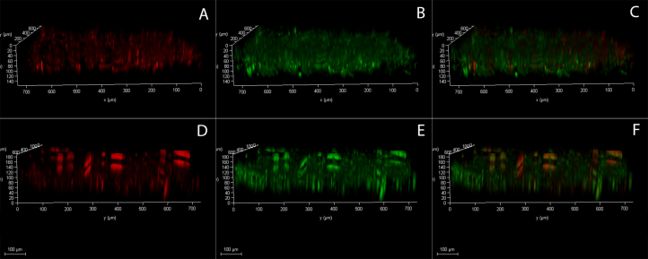
Filamentous fungi are ubiquitous and frequent components of biofilms. A means to visualize them and quantify their viability is essential for understanding their development and disruption. However, quantifying filamentous fungal biofilms poses challenges because, unlike yeasts and bacteria, they are not composed of discrete cells of similar size. This research focused on filamentous fungal biofilms that are representative of those in the built environment. The objective of this study was to develop a rapid method to examine biofilm structure and quantify live (metabolically active/ membrane undamaged) and dead (inactive/ membrane damaged) cells in biofilms utilizing a fluorescent probe staining method and confocal laser scanning microscopy (CLSM). For this, we compared two commercially available probe staining kits that have been developed for bacterial and yeast systems. One method utilized the classic cell stain FUN 1 that exhibits orange-red fluorescent intravacuolar structures in metabolically active cells, while dead cells are fluoresced green. The second method utilized a combination of SYTO9 and propidium iodide (PI), and stains cells based on their membrane morphology. SYTO9 is a green fluorescent stain with the capacity to penetrate the living cell walls, and PI is a red fluorescent stain that can only penetrate dead or dying cells with damaged cell membranes. Following staining, the biofilms were imaged using CLSM and biofilm volumes and thickness were quantified using COMSTAT, a computer program that measures biofilm accumulation from digital image stacks. The results were compared to independent measurements of live-dead cell density, as well as a classic cell viability assay-XTT. The data showed that the combination of SYTO9 and PI is optimal for staining filamentous fungal biofilms.

摘要

丝状真菌是生物膜中普遍且常见的组成部分。一种可视化它们并量化其活力的方法对于理解其发育和破坏至关重要。然而,量化丝状真菌生物膜存在挑战,因为与酵母和细菌不同,它们不是由大小相似的离散细胞组成。本研究聚焦于建筑环境中具有代表性的丝状真菌生物膜。本研究的目的是开发一种快速方法,利用荧光探针染色法和共聚焦激光扫描显微镜(CLSM)来检查生物膜结构并量化生物膜中活(代谢活跃/膜未受损)细胞和死(无活性/膜受损)细胞。为此,我们比较了两种为细菌和酵母系统开发的市售探针染色试剂盒。一种方法使用经典的细胞染料FUN 1,其在代谢活跃的细胞中呈现橙红色荧光液泡内结构,而死细胞发绿色荧光。第二种方法使用SYTO9和碘化丙啶(PI)的组合,并根据细胞膜形态对细胞进行染色。SYTO9是一种绿色荧光染料,能够穿透活细胞壁,而PI是一种红色荧光染料,只能穿透细胞膜受损的死细胞或濒死细胞。染色后,使用CLSM对生物膜进行成像,并使用COMSTAT(一种从数字图像堆栈测量生物膜积累的计算机程序)对生物膜体积和厚度进行量化。将结果与活死细胞密度的独立测量结果以及经典的细胞活力测定-XTT进行比较。数据表明,SYTO9和PI的组合对于染色丝状真菌生物膜是最佳的。

https://cdn.ncbi.nlm.nih.gov/pmc/blobs/2d7a/9646680/3f504ddbb55f/gr7.jpg
https://cdn.ncbi.nlm.nih.gov/pmc/blobs/2d7a/9646680/29322c811fbb/gr1.jpg
https://cdn.ncbi.nlm.nih.gov/pmc/blobs/2d7a/9646680/588b9d67f379/gr2.jpg
https://cdn.ncbi.nlm.nih.gov/pmc/blobs/2d7a/9646680/f64e447a6cc9/gr3.jpg
https://cdn.ncbi.nlm.nih.gov/pmc/blobs/2d7a/9646680/8715876898c2/gr4.jpg
https://cdn.ncbi.nlm.nih.gov/pmc/blobs/2d7a/9646680/abcb6d643e0c/gr5.jpg
https://cdn.ncbi.nlm.nih.gov/pmc/blobs/2d7a/9646680/e96fb26fc2f2/gr6.jpg
https://cdn.ncbi.nlm.nih.gov/pmc/blobs/2d7a/9646680/3f504ddbb55f/gr7.jpg

相似文献

1
Comparison of cell viability assessment and visualization of biofilm with two fluorescent probe staining methods.两种荧光探针染色方法对生物膜细胞活力评估及可视化的比较
Biofilm. 2022 Oct 28;4:100090. doi: 10.1016/j.bioflm.2022.100090. eCollection 2022 Dec.
2
Green fluorescent protein-propidium iodide (GFP-PI) based assay for flow cytometric measurement of bacterial viability.基于绿色荧光蛋白-碘化丙啶(GFP-PI)的流式细胞术检测细菌活力的方法。
Cytometry A. 2004 Aug;60(2):165-72. doi: 10.1002/cyto.a.20026.
3
Detection of microscopic filamentous fungal biofilms - Choosing the suitable methodology.微观丝状真菌生物膜的检测——选择合适的方法
J Microbiol Methods. 2023 Feb;205:106676. doi: 10.1016/j.mimet.2023.106676. Epub 2023 Jan 21.
4
The use of new probes and stains for improved assessment of cell viability and extracellular polymeric substances in Candida albicans biofilms.使用新型探针和染色剂以改进对白色念珠菌生物膜中细胞活力和胞外聚合物的评估。
Mycopathologia. 2005 Apr;159(3):353-60. doi: 10.1007/s11046-004-6987-7.
5
Propidium iodide staining underestimates viability of adherent bacterial cells.碘化丙啶染色低估了贴壁细菌细胞的活力。
Sci Rep. 2019 Apr 24;9(1):6483. doi: 10.1038/s41598-019-42906-3.
6
Critical aspects of using bacterial cell viability assays with the fluorophores SYTO9 and propidium iodide.使用荧光染料SYTO9和碘化丙啶进行细菌细胞活力测定的关键方面。
BMC Microbiol. 2015 Feb 18;15:36. doi: 10.1186/s12866-015-0376-x.
7
Aspergillus fumigatus biofilm on primary human sinonasal epithelial culture.烟曲霉生物膜在原代人鼻腔鼻窦上皮细胞培养物上的生长。
Am J Rhinol Allergy. 2011 Jul-Aug;25(4):219-25. doi: 10.2500/ajra.2011.25.3622.
8
Quantification, Viability Assessment, and Visualization Strategies for Acinetobacter Biofilms.不动杆菌生物膜的量化、生存力评估及可视化策略。
J Vis Exp. 2023 Aug 4(198). doi: 10.3791/65517.
9
Artificial Pseudomonas aeruginosa biofilms and confocal laser scanning microscopic analysis.人工铜绿假单胞菌生物膜与共聚焦激光扫描显微镜分析
J Infect Chemother. 2001 Jun;7(2):87-93. doi: 10.1007/s101560100014.
10
Confocal laser scanning microscopy analysis of S. epidermidis biofilms exposed to farnesol, vancomycin and rifampicin.对暴露于法尼醇、万古霉素和利福平的表皮葡萄球菌生物膜进行共聚焦激光扫描显微镜分析。
BMC Res Notes. 2012 May 16;5:244. doi: 10.1186/1756-0500-5-244.

引用本文的文献

1
The Metabolic and Physiological Responses to Spaceflight of a Lipopeptide-Producing Bacillus subtilis.产脂肽枯草芽孢杆菌对太空飞行的代谢和生理反应
Microb Biotechnol. 2025 Mar;18(3):e70111. doi: 10.1111/1751-7915.70111.
2
Primary and Re-exposure effects of D-enantiomeric peptide on metabolism, diversity, and composition of oral biofilms at different stages of recovery.D-对映体肽对不同恢复阶段口腔生物膜代谢、多样性和组成的初次及再暴露效应。
Bioact Mater. 2025 Feb 20;48:257-272. doi: 10.1016/j.bioactmat.2025.02.023. eCollection 2025 Jun.
3
Membrane Damage and Metabolic Disruption as the Mechanisms of Linalool against : An Amino Acid Metabolomics Study.

本文引用的文献

1
Anti-microbial and anti-biofilm activities of combined chelerythrine-sanguinarine and mode of action against Candida albicans and Cryptococcus neoformans in vitro.联合白屈菜红碱-血根碱的抗微生物和抗生物膜活性及其对体外白色念珠菌和新型隐球菌的作用模式。
Colloids Surf B Biointerfaces. 2020 Jul;191:111003. doi: 10.1016/j.colsurfb.2020.111003. Epub 2020 Apr 1.
2
In vitro Antifungal Effects of Berberine Against spp. In Planktonic and Biofilm Conditions.小檗碱在浮游和生物膜条件下对 种真菌的体外抗真菌作用。 (注:原文中“ spp.”部分表述不完整,可能影响准确理解)
Drug Des Devel Ther. 2020 Jan 9;14:87-101. doi: 10.2147/DDDT.S230857. eCollection 2020.
3
膜损伤和代谢紊乱作为芳樟醇作用机制的研究:一项氨基酸代谢组学研究
Foods. 2024 Aug 9;13(16):2501. doi: 10.3390/foods13162501.
4
Large-Scale Isolation of Milk Exosomes for Skincare.用于护肤品的大规模牛奶外泌体分离
Pharmaceutics. 2024 Jul 11;16(7):930. doi: 10.3390/pharmaceutics16070930.
5
Improving the biocompatibility and antibacterial efficacy of silver nanoparticles functionalized with (LLRR) antimicrobial peptide.用(LLRR)抗菌肽功能化提高银纳米粒子的生物相容性和抗菌功效。
World J Microbiol Biotechnol. 2023 Nov 4;40(1):1. doi: 10.1007/s11274-023-03792-0.
6
Metabolomic Differences between Viable but Nonculturable and Recovered Zhang.活的但不可培养状态与复苏状态的张之间的代谢组学差异
Foods. 2023 Sep 18;12(18):3472. doi: 10.3390/foods12183472.
7
Dynamic full-field optical coherence tomography for live-cell imaging and growth-phase monitoring in .动态全场光学相干断层扫描用于. 活细胞成像和生长阶段监测。
Front Cell Infect Microbiol. 2023 Jul 12;13:1183340. doi: 10.3389/fcimb.2023.1183340. eCollection 2023.
Developmental stages of biofilm and characterization of extracellular matrix of manglicolous fungus Aspergillus niger BSC-1.
黑曲霉 BSC-1 生物膜发育阶段及胞外基质特性研究
J Basic Microbiol. 2020 Mar;60(3):231-242. doi: 10.1002/jobm.201900550. Epub 2019 Dec 19.
4
Biofilm characterization of Fusarium solani keratitis isolate: increased resistance to antifungals and UV light.茄病镰刀菌角膜炎分离株的生物膜特性:对抗真菌药物和紫外线的抵抗力增加。
J Microbiol. 2019 Jun;57(6):485-497. doi: 10.1007/s12275-019-8637-2. Epub 2019 May 27.
5
Propidium iodide staining underestimates viability of adherent bacterial cells.碘化丙啶染色低估了贴壁细菌细胞的活力。
Sci Rep. 2019 Apr 24;9(1):6483. doi: 10.1038/s41598-019-42906-3.
6
Antimicrobial properties of a new type of photosensitizer derived from phthalocyanine against planktonic and biofilm forms of Staphylococcus aureus.新型酞菁类光敏剂抗浮游和生物膜形式金黄色葡萄球菌的抗菌性能。
Photodiagnosis Photodyn Ther. 2018 Mar;21:316-326. doi: 10.1016/j.pdpdt.2018.01.003. Epub 2018 Jan 4.
7
Analysis and description of the stages of Aspergillus fumigatus biofilm formation using scanning electron microscopy.使用扫描电子显微镜对烟曲霉生物膜形成阶段的分析与描述。
BMC Microbiol. 2016 Oct 18;16(1):243. doi: 10.1186/s12866-016-0859-4.
8
Critical aspects of using bacterial cell viability assays with the fluorophores SYTO9 and propidium iodide.使用荧光染料SYTO9和碘化丙啶进行细菌细胞活力测定的关键方面。
BMC Microbiol. 2015 Feb 18;15:36. doi: 10.1186/s12866-015-0376-x.
9
Molecular mechanisms of antimicrobial tolerance and resistance in bacterial and fungal biofilms.细菌和真菌生物膜中抗菌药物耐受和耐药的分子机制。
Trends Microbiol. 2014 Jun;22(6):326-33. doi: 10.1016/j.tim.2014.02.001. Epub 2014 Mar 2.
10
The role of bacterial biofilms and surface components in plant-bacterial associations.细菌生物膜和表面成分在植物-细菌共生关系中的作用。
Int J Mol Sci. 2013 Jul 30;14(8):15838-59. doi: 10.3390/ijms140815838.