• 文献检索
  • 文档翻译
  • 深度研究
  • 学术资讯
  • Suppr Zotero 插件Zotero 插件
  • 邀请有礼
  • 套餐&价格
  • 历史记录
应用&插件
Suppr Zotero 插件Zotero 插件浏览器插件Mac 客户端Windows 客户端微信小程序
定价
高级版会员购买积分包购买API积分包
服务
文献检索文档翻译深度研究API 文档MCP 服务
关于我们
关于 Suppr公司介绍联系我们用户协议隐私条款
关注我们

Suppr 超能文献

核心技术专利:CN118964589B侵权必究
粤ICP备2023148730 号-1Suppr @ 2026

文献检索

告别复杂PubMed语法,用中文像聊天一样搜索,搜遍4000万医学文献。AI智能推荐,让科研检索更轻松。

立即免费搜索

文件翻译

保留排版,准确专业,支持PDF/Word/PPT等文件格式,支持 12+语言互译。

免费翻译文档

深度研究

AI帮你快速写综述,25分钟生成高质量综述,智能提取关键信息,辅助科研写作。

立即免费体验

纵向在体钙成像揭示了单细胞水平上病变视网膜神经节细胞的动态活动变化。

Longitudinal in vivo Ca imaging reveals dynamic activity changes of diseased retinal ganglion cells at the single-cell level.

机构信息

Spencer Center for Vision Research, Department of Ophthalmology, Byers Eye Institute at Stanford University School of Medicine, Palo Alto, CA 94304.

Department of Ophthalmology, The Second Xiangya Hospital, Central South University, Changsha 410011, China.

出版信息

Proc Natl Acad Sci U S A. 2022 Nov 29;119(48):e2206829119. doi: 10.1073/pnas.2206829119. Epub 2022 Nov 21.

DOI:10.1073/pnas.2206829119
PMID:36409915
原文链接:https://pmc.ncbi.nlm.nih.gov/articles/PMC9889883/
Abstract

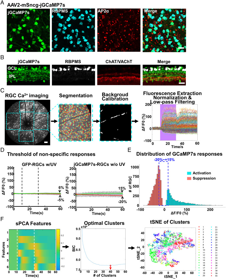
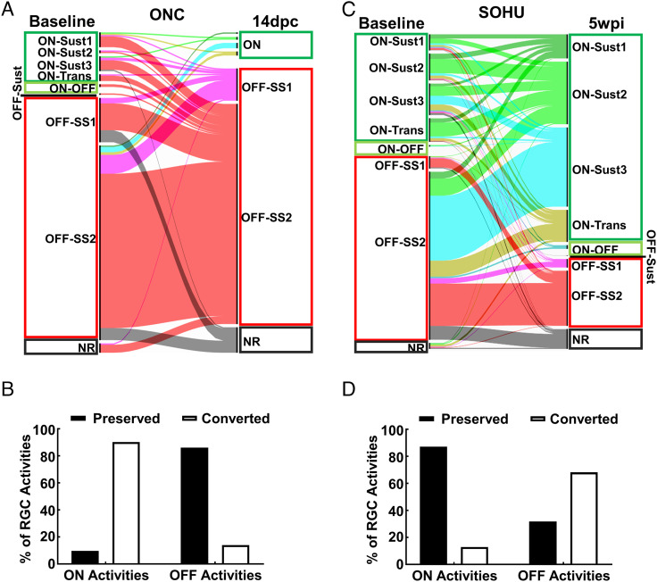
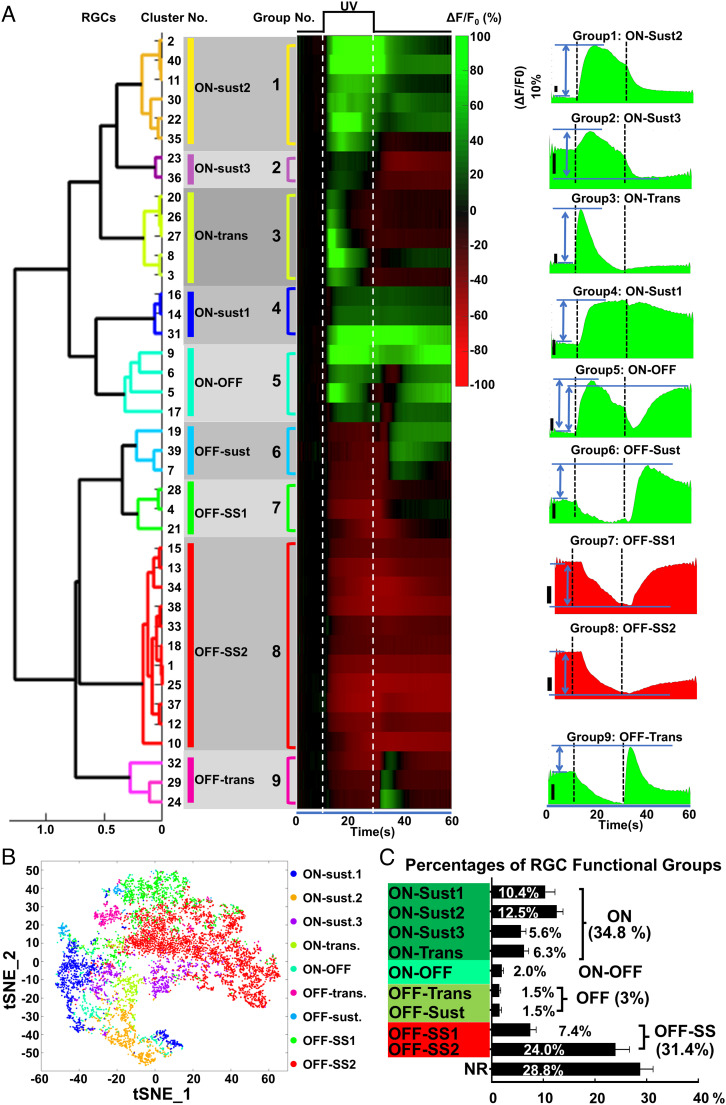
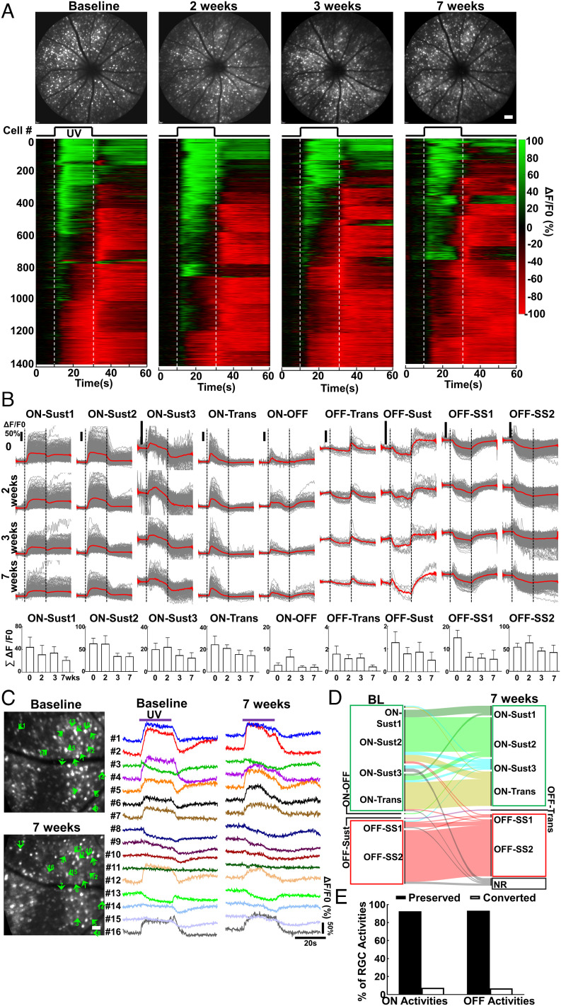
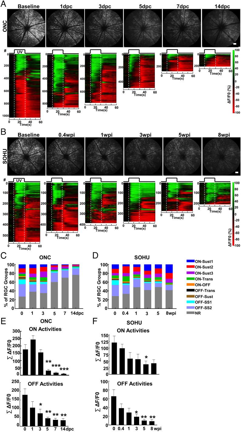
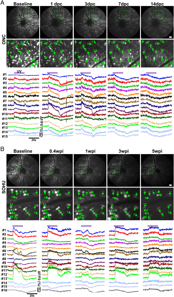
Retinal ganglion cells (RGCs) are heterogeneous projection neurons that convey distinct visual features from the retina to brain. Here, we present a high-throughput in vivo RGC activity assay in response to light stimulation using noninvasive Ca imaging of thousands of RGCs simultaneously in living mice. Population and single-cell analyses of longitudinal RGC Ca imaging reveal distinct functional responses of RGCs and unprecedented individual RGC activity conversions during traumatic and glaucomatous degeneration. This study establishes a foundation for future in vivo RGC function classifications and longitudinal activity evaluations using more advanced imaging techniques and visual stimuli under normal, disease, and neural repair conditions. These analyses can be performed at both the population and single-cell levels using temporal and spatial information, which will be invaluable for understanding RGC pathophysiology and identifying functional biomarkers for diverse optic neuropathies.

摘要

视网膜神经节细胞(RGCs)是一种异质性的投射神经元,它们将不同的视觉特征从视网膜传递到大脑。在这里,我们使用活体小鼠中数千个 RGC 的非侵入性 Ca 成像,展示了一种针对光刺激的高通量体内 RGC 活性测定方法。对纵向 RGC Ca 成像的群体和单细胞分析揭示了 RGC 的不同功能反应,以及在创伤性和青光眼变性过程中前所未有的个体 RGC 活性转换。这项研究为未来使用更先进的成像技术和视觉刺激在正常、疾病和神经修复条件下进行体内 RGC 功能分类和纵向活性评估奠定了基础。这些分析可以在群体和单细胞水平上使用时间和空间信息来进行,这对于理解 RGC 的病理生理学和确定各种视神经病变的功能生物标志物将是非常有价值的。

https://cdn.ncbi.nlm.nih.gov/pmc/blobs/0b98/9889883/1249c63e79ba/pnas.2206829119fig06.jpg
https://cdn.ncbi.nlm.nih.gov/pmc/blobs/0b98/9889883/19b07467c49f/pnas.2206829119fig01.jpg
https://cdn.ncbi.nlm.nih.gov/pmc/blobs/0b98/9889883/0eceb2aab056/pnas.2206829119fig02.jpg
https://cdn.ncbi.nlm.nih.gov/pmc/blobs/0b98/9889883/eb2765d16b1e/pnas.2206829119fig03.jpg
https://cdn.ncbi.nlm.nih.gov/pmc/blobs/0b98/9889883/5124a9e75843/pnas.2206829119fig04.jpg
https://cdn.ncbi.nlm.nih.gov/pmc/blobs/0b98/9889883/44b8535bd378/pnas.2206829119fig05.jpg
https://cdn.ncbi.nlm.nih.gov/pmc/blobs/0b98/9889883/1249c63e79ba/pnas.2206829119fig06.jpg

相似文献

1
Longitudinal in vivo Ca imaging reveals dynamic activity changes of diseased retinal ganglion cells at the single-cell level.纵向在体钙成像揭示了单细胞水平上病变视网膜神经节细胞的动态活动变化。
Proc Natl Acad Sci U S A. 2022 Nov 29;119(48):e2206829119. doi: 10.1073/pnas.2206829119. Epub 2022 Nov 21.
2
Reconstruction of natural images from responses of primate retinal ganglion cells.从灵长类视网膜神经节细胞的反应中重建自然图像。
Elife. 2020 Nov 4;9:e58516. doi: 10.7554/eLife.58516.
3
Strip1 regulates retinal ganglion cell survival by suppressing Jun-mediated apoptosis to promote retinal neural circuit formation.Strip1 通过抑制 Jun 介导的细胞凋亡来调节视网膜神经节细胞的存活,从而促进视网膜神经回路的形成。
Elife. 2022 Mar 22;11:e74650. doi: 10.7554/eLife.74650.
4
Assessment of inner retina dysfunction and progressive ganglion cell loss in a mouse model of glaucoma.评估青光眼小鼠模型中内视网膜功能障碍和节细胞进行性丧失。
Exp Eye Res. 2014 May;122:40-9. doi: 10.1016/j.exer.2014.02.022. Epub 2014 Mar 12.
5
Establishing the ground squirrel as a superb model for retinal ganglion cell disorders and optic neuropathies.确立地松鼠为视网膜神经节细胞病变和视神经病变的优秀模型。
Lab Invest. 2021 Sep;101(9):1289-1303. doi: 10.1038/s41374-021-00637-y. Epub 2021 Jul 12.
6
A freely available semi-automated method for quantifying retinal ganglion cells in entire retinal flatmounts.一种可免费使用的半自动方法,用于对整个视网膜平铺标本中的视网膜神经节细胞进行定量分析。
Exp Eye Res. 2016 Jun;147:105-113. doi: 10.1016/j.exer.2016.04.010. Epub 2016 Apr 20.
7
Mild Intraocular Pressure Elevation in Mice Reveals Distinct Retinal Ganglion Cell Functional Thresholds and Pressure-Dependent Properties.轻度眼内压升高揭示了小鼠视网膜神经节细胞功能的不同阈值和压力依赖性特征。
J Neurosci. 2019 Mar 6;39(10):1881-1891. doi: 10.1523/JNEUROSCI.2085-18.2019. Epub 2019 Jan 8.
8
Bax Contributes to Retinal Ganglion Cell Dendritic Degeneration During Glaucoma.Bax 促进青光眼期间视网膜神经节细胞树突退化。
Mol Neurobiol. 2022 Mar;59(3):1366-1380. doi: 10.1007/s12035-021-02675-5. Epub 2022 Jan 5.
9
Effects of acute delivery of endothelin-1 on retinal ganglion cell loss in the rat.急性给予内皮素-1对大鼠视网膜神经节细胞丢失的影响。
Exp Eye Res. 2006 Jan;82(1):132-45. doi: 10.1016/j.exer.2005.06.002. Epub 2005 Jul 19.
10
Neuritin 1 Drives Therapeutic Preservation of Retinal Ganglion Cells in an Human Glaucoma Model.神经突蛋白1在人青光眼模型中驱动视网膜神经节细胞的治疗性保护。
J Ocul Pharmacol Ther. 2024 Nov;40(9):596-607. doi: 10.1089/jop.2024.0041. Epub 2024 Jul 12.

引用本文的文献

1
Effect of brimonidine on retinal ganglion cell function by in vivo calcium imaging of optic nerve crush in mice.溴莫尼定对小鼠视神经挤压伤后视网膜神经节细胞功能的影响:通过活体钙成像技术进行研究
Exp Eye Res. 2025 Jun;255:110355. doi: 10.1016/j.exer.2025.110355. Epub 2025 Mar 22.
2
Damage and repair in retinal degenerative diseases: Molecular basis through clinical translation.视网膜退行性疾病中的损伤与修复:从分子基础到临床转化
Neural Regen Res. 2026 Apr 1;21(4):1383-1395. doi: 10.4103/NRR.NRR-D-24-01016. Epub 2025 Feb 24.
3
Endoplasmic reticulum stress-related deficits in calcium clearance promote neuronal dysfunction that is prevented by SERCA2 gene augmentation.

本文引用的文献

1
An uncommon neuronal class conveys visual signals from rods and cones to retinal ganglion cells.一种不常见的神经元细胞类群将视觉信号从视杆细胞和视锥细胞传递到视网膜神经节细胞。
Proc Natl Acad Sci U S A. 2021 Nov 2;118(44). doi: 10.1073/pnas.2104884118.
2
Chronic mild and acute severe glaucomatous neurodegeneration derived from silicone oil-induced ocular hypertension.慢性轻度和急性重度青光眼性神经退行性变源于硅油诱导的眼内高压。
Sci Rep. 2021 Apr 27;11(1):9052. doi: 10.1038/s41598-021-88690-x.
3
Commonalities of optic nerve injury and glaucoma-induced neurodegeneration: Insights from transcriptome-wide studies.
内质网应激相关的钙清除缺陷会促进神经元功能障碍,而SERCA2基因增强可预防这种功能障碍。
Cell Rep Med. 2024 Dec 17;5(12):101839. doi: 10.1016/j.xcrm.2024.101839. Epub 2024 Nov 29.
4
A novel GCaMP6f-RCS rat model for studying electrical stimulation in the degenerated retina.一种用于研究退化视网膜电刺激的新型GCaMP6f-RCS大鼠模型。
Front Cell Dev Biol. 2024 Apr 22;12:1386141. doi: 10.3389/fcell.2024.1386141. eCollection 2024.
5
Osteopontin drives retinal ganglion cell resiliency in glaucomatous optic neuropathy.骨桥蛋白驱动青光眼视神经病变中视网膜神经节细胞的弹性。
Cell Rep. 2023 Sep 26;42(9):113038. doi: 10.1016/j.celrep.2023.113038. Epub 2023 Aug 23.
6
Retinal microvascular and neuronal pathologies probed in vivo by adaptive optical two-photon fluorescence microscopy.自适应光学双光子荧光显微镜在体探测视网膜微血管和神经元病变。
Elife. 2023 Apr 11;12:e84853. doi: 10.7554/eLife.84853.
7
The importance of unambiguous cell origin determination in neuronal repopulation studies.在神经元再填充研究中明确确定细胞来源的重要性。
iScience. 2023 Mar 9;26(4):106361. doi: 10.1016/j.isci.2023.106361. eCollection 2023 Apr 21.
视神经损伤与青光眼相关性神经退行性变的共性:来自转录组范围研究的启示。
Exp Eye Res. 2021 Jun;207:108571. doi: 10.1016/j.exer.2021.108571. Epub 2021 Apr 15.
4
Adaptive responses to neurodegenerative stress in glaucoma.青光眼神经退行性应激的适应性反应。
Prog Retin Eye Res. 2021 Sep;84:100953. doi: 10.1016/j.preteyeres.2021.100953. Epub 2021 Feb 25.
5
In vivo evaluation of retinal ganglion cells and optic nerve's integrity in large animals by multi-modality analysis.多模态分析评估大动物视网膜神经节细胞和视神经的完整性。
Exp Eye Res. 2020 Aug;197:108117. doi: 10.1016/j.exer.2020.108117. Epub 2020 Jun 27.
6
True S-cones are concentrated in the ventral mouse retina and wired for color detection in the upper visual field.真 S 视锥细胞集中在老鼠视网膜的腹侧,并且在上半视野中用于颜色检测。
Elife. 2020 May 28;9:e56840. doi: 10.7554/eLife.56840.
7
Adaptive optics two-photon microscopy enables near-diffraction-limited and functional retinal imaging in vivo.自适应光学双光子显微镜能够在体内实现近衍射极限的功能性视网膜成像。
Light Sci Appl. 2020 May 6;9:79. doi: 10.1038/s41377-020-0317-9. eCollection 2020.
8
Longitudinal Morphological and Functional Assessment of RGC Neurodegeneration After Optic Nerve Crush in Mouse.小鼠视神经挤压后视网膜神经节细胞神经变性的纵向形态学和功能评估
Front Cell Neurosci. 2020 Apr 29;14:109. doi: 10.3389/fncel.2020.00109. eCollection 2020.
9
Mouse γ-Synuclein Promoter-Mediated Gene Expression and Editing in Mammalian Retinal Ganglion Cells.鼠 γ-突触核蛋白启动子介导的哺乳动物视网膜神经节细胞中的基因表达和编辑。
J Neurosci. 2020 May 13;40(20):3896-3914. doi: 10.1523/JNEUROSCI.0102-20.2020. Epub 2020 Apr 16.
10
Fundamental bounds on the fidelity of sensory cortical coding.感觉皮层编码保真度的基本界限。
Nature. 2020 Apr;580(7801):100-105. doi: 10.1038/s41586-020-2130-2. Epub 2020 Mar 18.