• 文献检索
  • 文档翻译
  • 深度研究
  • 学术资讯
  • Suppr Zotero 插件Zotero 插件
  • 邀请有礼
  • 套餐&价格
  • 历史记录
应用&插件
Suppr Zotero 插件Zotero 插件浏览器插件Mac 客户端Windows 客户端微信小程序
定价
高级版会员购买积分包购买API积分包
服务
文献检索文档翻译深度研究API 文档MCP 服务
关于我们
关于 Suppr公司介绍联系我们用户协议隐私条款
关注我们

Suppr 超能文献

核心技术专利:CN118964589B侵权必究
粤ICP备2023148730 号-1Suppr @ 2026

文献检索

告别复杂PubMed语法,用中文像聊天一样搜索,搜遍4000万医学文献。AI智能推荐,让科研检索更轻松。

立即免费搜索

文件翻译

保留排版,准确专业,支持PDF/Word/PPT等文件格式,支持 12+语言互译。

免费翻译文档

深度研究

AI帮你快速写综述,25分钟生成高质量综述,智能提取关键信息,辅助科研写作。

立即免费体验

骨桥蛋白驱动青光眼视神经病变中视网膜神经节细胞的弹性。

Osteopontin drives retinal ganglion cell resiliency in glaucomatous optic neuropathy.

机构信息

Department of Ophthalmology, University of California San Francisco, San Francisco, CA 94158, USA.

Department of Ophthalmology, University of California San Francisco, San Francisco, CA 94158, USA.

出版信息

Cell Rep. 2023 Sep 26;42(9):113038. doi: 10.1016/j.celrep.2023.113038. Epub 2023 Aug 23.

DOI:10.1016/j.celrep.2023.113038
PMID:37624696
原文链接:https://pmc.ncbi.nlm.nih.gov/articles/PMC10591811/
Abstract

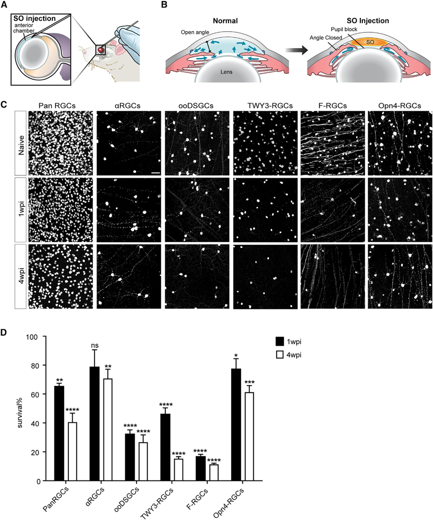
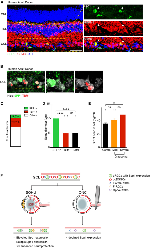
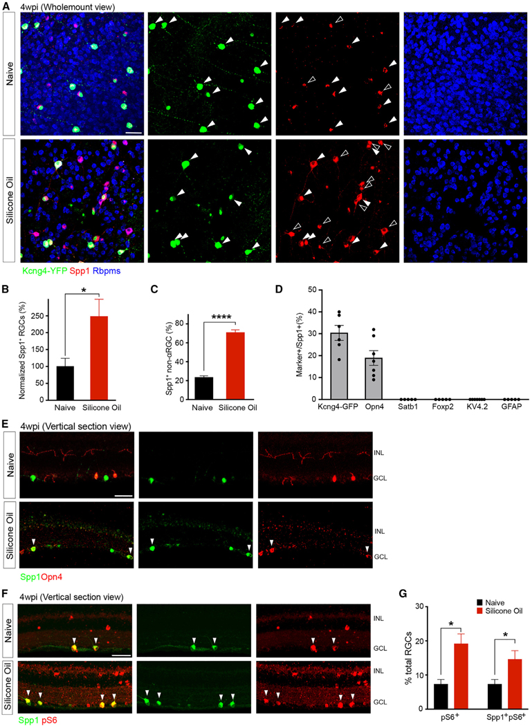
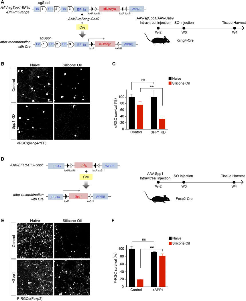
Chronic neurodegeneration and acute injuries lead to neuron losses via diverse processes. We compared retinal ganglion cell (RGC) responses between chronic glaucomatous conditions and the acute injury model. Among major RGC subclasses, αRGCs and intrinsically photosensitive RGCs (ipRGCs) preferentially survive glaucomatous conditions, similar to findings in the retina subject to axotomy. Focusing on an αRGC intrinsic factor, Osteopontin (secreted phosphoprotein 1 [Spp1]), we found an ectopic neuronal expression of Osteopontin (Spp1) in other RGCs subject to glaucomatous conditions. This contrasted with the Spp1 downregulation subject to axotomy. αRGC-specific Spp1 elimination led to significant αRGC loss, diminishing their resiliency. Spp1 overexpression led to robust neuroprotection of susceptible RGC subclasses under glaucomatous conditions. In contrast, Spp1 overexpression did not significantly protect RGCs subject to axotomy. Additionally, SPP1 marked adult human RGC subsets with large somata and SPP1 expression in the aqueous humor correlated with glaucoma severity. Our study reveals Spp1's role in mediating neuronal resiliency in glaucoma.

摘要

慢性神经退行性变和急性损伤通过不同的过程导致神经元丢失。我们比较了慢性青光眼状态和急性损伤模型之间的视网膜神经节细胞 (RGC) 反应。在主要的 RGC 亚类中,αRGC 和内在光敏感 RGC (ipRGC) 优先在青光眼条件下存活,这与在经历轴突切断的视网膜中的发现相似。我们关注αRGC 的内在因素骨桥蛋白 (分泌磷蛋白 1[Spp1]),发现在青光眼条件下,其他 RGC 中存在骨桥蛋白 (Spp1) 的异位神经元表达。这与轴突切断时 Spp1 的下调形成对比。αRGC 特异性 Spp1 消除导致显著的 αRGC 丢失,降低了它们的弹性。Spp1 的过表达导致易感 RGC 亚类在青光眼条件下的强大神经保护作用。相比之下,Spp1 的过表达并不能显著保护经历轴突切断的 RGC。此外,SPP1 标记了具有大胞体的成年人类 RGC 亚群,房水中的 SPP1 表达与青光眼的严重程度相关。我们的研究揭示了 Spp1 在介导青光眼神经元弹性中的作用。

https://cdn.ncbi.nlm.nih.gov/pmc/blobs/61b5/10591811/4fc503b56aeb/nihms-1933884-f0004.jpg
https://cdn.ncbi.nlm.nih.gov/pmc/blobs/61b5/10591811/9e1813f64d45/nihms-1933884-f0001.jpg
https://cdn.ncbi.nlm.nih.gov/pmc/blobs/61b5/10591811/0b4c08f62022/nihms-1933884-f0002.jpg
https://cdn.ncbi.nlm.nih.gov/pmc/blobs/61b5/10591811/f0b2b149e868/nihms-1933884-f0003.jpg
https://cdn.ncbi.nlm.nih.gov/pmc/blobs/61b5/10591811/4fc503b56aeb/nihms-1933884-f0004.jpg

相似文献

1
Osteopontin drives retinal ganglion cell resiliency in glaucomatous optic neuropathy.骨桥蛋白驱动青光眼视神经病变中视网膜神经节细胞的弹性。
Cell Rep. 2023 Sep 26;42(9):113038. doi: 10.1016/j.celrep.2023.113038. Epub 2023 Aug 23.
2
Sphingosine 1-phosphate receptor 1 is required for retinal ganglion cell survival after optic nerve trauma.1-磷酸鞘氨醇受体1是视神经损伤后视网膜神经节细胞存活所必需的。
J Neurochem. 2016 Aug;138(4):571-86. doi: 10.1111/jnc.13701. Epub 2016 Jul 6.
3
Secreted phosphoprotein 1 slows neurodegeneration and rescues visual function in mouse models of aging and glaucoma.分泌性磷蛋白 1 可减缓衰老和青光眼小鼠模型的神经退行性变并挽救其视觉功能。
Cell Rep. 2022 Dec 27;41(13):111880. doi: 10.1016/j.celrep.2022.111880.
4
Survival of Alpha and Intrinsically Photosensitive Retinal Ganglion Cells in NMDA-Induced Neurotoxicity and a Mouse Model of Normal Tension Glaucoma.NMDA 诱导的神经毒性和正常眼压型青光眼小鼠模型中 Alpha 和内在光敏性视网膜神经节细胞的存活。
Invest Ophthalmol Vis Sci. 2019 Sep 3;60(12):3696-3707. doi: 10.1167/iovs.19-27145.
5
An agonistic TrkB mAb causes sustained TrkB activation, delays RGC death, and protects the retinal structure in optic nerve axotomy and in glaucoma.一种激动型 TrkB mAb 可引起持续的 TrkB 激活,延迟 RGC 死亡,并在视神经切断和青光眼模型中保护视网膜结构。
Invest Ophthalmol Vis Sci. 2010 Sep;51(9):4722-31. doi: 10.1167/iovs.09-5032. Epub 2010 Mar 31.
6
Down-regulation of semaphorin 3F in rat retinal ganglion cells in response to optic nerve crush.大鼠视网膜神经节细胞中信号素3F在视神经挤压后下调。
Cell Biochem Funct. 2016 Jul;34(5):378-84. doi: 10.1002/cbf.3200. Epub 2016 Jun 16.
7
Protection of mouse retinal ganglion cell axons and soma from glaucomatous and ischemic injury by cytoplasmic overexpression of Nmnat1.通过细胞质过表达 Nmnat1 保护小鼠视网膜神经节细胞轴突和体免受青光眼和缺血性损伤。
Invest Ophthalmol Vis Sci. 2013 Jan 2;54(1):25-36. doi: 10.1167/iovs.12-10861.
8
Functional analysis of mesencephalic astrocyte-derived neurotrophic factor in retinal ganglion cells under oxidative stress.在氧化应激下中脑星形胶质细胞衍生的神经营养因子对视网膜神经节细胞的功能分析。
Cell Biochem Funct. 2021 Jan;39(1):98-106. doi: 10.1002/cbf.3567. Epub 2020 Jul 1.
9
Intravitreal delivery of human NgR-Fc decoy protein regenerates axons after optic nerve crush and protects ganglion cells in glaucoma models.玻璃体内注射人NgR-Fc诱饵蛋白可使视神经挤压伤后轴突再生,并在青光眼模型中保护神经节细胞。
Invest Ophthalmol Vis Sci. 2015 Feb 5;56(2):1357-66. doi: 10.1167/iovs.14-15472.
10
Role of hypoxia-inducible factor-1α in preconditioning-induced protection of retinal ganglion cells in glaucoma.缺氧诱导因子-1α在青光眼预处理诱导的视网膜神经节细胞保护中的作用
Mol Vis. 2013 Nov 23;19:2360-72. eCollection 2013.

引用本文的文献

1
The pivotal role of inflammatory factors in glaucoma: a systematic review.炎症因子在青光眼发病机制中的关键作用:一项系统综述
Front Immunol. 2025 May 23;16:1577200. doi: 10.3389/fimmu.2025.1577200. eCollection 2025.
2
Molecular mechanisms after optic nerve injury: Neurorepair strategies from a transcriptomic perspective.视神经损伤后的分子机制:从转录组学角度看神经修复策略。
Neural Regen Res. 2025 Apr 29. doi: 10.4103/NRR.NRR-D-24-00794.
3
Optic nerve injury impairs intrinsic mechanisms underlying electrical activity in a resilient retinal ganglion cell.

本文引用的文献

1
Secreted phosphoprotein 1 slows neurodegeneration and rescues visual function in mouse models of aging and glaucoma.分泌性磷蛋白 1 可减缓衰老和青光眼小鼠模型的神经退行性变并挽救其视觉功能。
Cell Rep. 2022 Dec 27;41(13):111880. doi: 10.1016/j.celrep.2022.111880.
2
Longitudinal in vivo Ca imaging reveals dynamic activity changes of diseased retinal ganglion cells at the single-cell level.纵向在体钙成像揭示了单细胞水平上病变视网膜神经节细胞的动态活动变化。
Proc Natl Acad Sci U S A. 2022 Nov 29;119(48):e2206829119. doi: 10.1073/pnas.2206829119. Epub 2022 Nov 21.
3
Cell atlas of the human ocular anterior segment: Tissue-specific and shared cell types.
视神经损伤会损害具有适应性的视网膜神经节细胞电活动的内在机制。
J Physiol. 2025 Feb 22. doi: 10.1113/JP286414.
4
Expression of Osteopontin in M2 and M4 Intrinsically Photosensitive Retinal Ganglion Cells in the Mouse Retina.骨桥蛋白在小鼠视网膜M2和M4内在光敏性视网膜神经节细胞中的表达
Invest Ophthalmol Vis Sci. 2025 Feb 3;66(2):14. doi: 10.1167/iovs.66.2.14.
5
Afadin Sorts Different Retinal Neuron Types into Accurate Cellular Layers.Afadin将不同类型的视网膜神经元分类到精确的细胞层中。
bioRxiv. 2024 Dec 25:2024.12.24.630272. doi: 10.1101/2024.12.24.630272.
6
Molecular and spatial analysis of ganglion cells on retinal flatmounts: diversity, topography, and perivascularity.视网膜铺片上神经节细胞的分子与空间分析:多样性、地形图及血管周特性
bioRxiv. 2024 Dec 17:2024.12.15.628587. doi: 10.1101/2024.12.15.628587.
7
Emerging strategies targeting genes and cells in glaucoma.青光眼治疗中针对基因和细胞的新兴策略。
Vision Res. 2025 Feb;227:108533. doi: 10.1016/j.visres.2024.108533. Epub 2024 Dec 6.
8
Monocyte Invasion into the Retina Restricts the Regeneration of Neurons from Müller Glia.单核细胞浸润视网膜限制了 Müller 胶质细胞来源神经元的再生。
J Neurosci. 2024 Nov 13;44(46):e0938242024. doi: 10.1523/JNEUROSCI.0938-24.2024.
9
Proteomics Analyses of Small Extracellular Vesicles of Aqueous Humor: Identification and Validation of GAS6 and SPP1 as Glaucoma Markers.水液中小细胞外囊泡的蛋白质组学分析:GAS6 和 SPP1 作为青光眼标志物的鉴定和验证。
Int J Mol Sci. 2024 Jun 26;25(13):6995. doi: 10.3390/ijms25136995.
10
An Update to Biomechanical and Biochemical Principles of Retinal Injury in Child Abuse.虐待儿童中视网膜损伤的生物力学和生物化学原理的更新
Children (Basel). 2024 May 12;11(5):586. doi: 10.3390/children11050586.
人类眼前节细胞图谱:组织特异性和共享细胞类型。
Proc Natl Acad Sci U S A. 2022 Jul 19;119(29):e2200914119. doi: 10.1073/pnas.2200914119. Epub 2022 Jul 15.
4
Core transcription programs controlling injury-induced neurodegeneration of retinal ganglion cells.核心转录程序控制视网膜神经节细胞损伤诱导的神经变性。
Neuron. 2022 Aug 17;110(16):2607-2624.e8. doi: 10.1016/j.neuron.2022.06.003. Epub 2022 Jun 28.
5
Trans-Seq maps a selective mammalian retinotectal synapse instructed by Nephronectin.Trans-Seq 图谱描绘了由 Nephrontin 指导的选择性哺乳动物视顶盖突触。
Nat Neurosci. 2022 May;25(5):659-674. doi: 10.1038/s41593-022-01068-8. Epub 2022 May 6.
6
Molecular characterization of selectively vulnerable neurons in Alzheimer's disease.阿尔茨海默病中选择性易损神经元的分子特征。
Nat Neurosci. 2021 Feb;24(2):276-287. doi: 10.1038/s41593-020-00764-7. Epub 2021 Jan 11.
7
Differential susceptibility of retinal ganglion cell subtypes in acute and chronic models of injury and disease.急性和慢性损伤及疾病模型中视网膜神经节细胞亚型的易感性差异。
Sci Rep. 2020 Oct 15;10(1):17359. doi: 10.1038/s41598-020-71460-6.
8
Axon Regeneration in the Mammalian Optic Nerve.哺乳动物视神经中的轴突再生。
Annu Rev Vis Sci. 2020 Sep 15;6:195-213. doi: 10.1146/annurev-vision-022720-094953.
9
Cell Atlas of The Human Fovea and Peripheral Retina.人眼黄斑和周边视网膜细胞图谱。
Sci Rep. 2020 Jun 17;10(1):9802. doi: 10.1038/s41598-020-66092-9.
10
Longitudinal Morphological and Functional Assessment of RGC Neurodegeneration After Optic Nerve Crush in Mouse.小鼠视神经挤压后视网膜神经节细胞神经变性的纵向形态学和功能评估
Front Cell Neurosci. 2020 Apr 29;14:109. doi: 10.3389/fncel.2020.00109. eCollection 2020.