• 文献检索
  • 文档翻译
  • 深度研究
  • 学术资讯
  • Suppr Zotero 插件Zotero 插件
  • 邀请有礼
  • 套餐&价格
  • 历史记录
应用&插件
Suppr Zotero 插件Zotero 插件浏览器插件Mac 客户端Windows 客户端微信小程序
定价
高级版会员购买积分包购买API积分包
服务
文献检索文档翻译深度研究API 文档MCP 服务
关于我们
关于 Suppr公司介绍联系我们用户协议隐私条款
关注我们

Suppr 超能文献

核心技术专利:CN118964589B侵权必究
粤ICP备2023148730 号-1Suppr @ 2026

文献检索

告别复杂PubMed语法,用中文像聊天一样搜索,搜遍4000万医学文献。AI智能推荐,让科研检索更轻松。

立即免费搜索

文件翻译

保留排版,准确专业,支持PDF/Word/PPT等文件格式,支持 12+语言互译。

免费翻译文档

深度研究

AI帮你快速写综述,25分钟生成高质量综述,智能提取关键信息,辅助科研写作。

立即免费体验

真 S 视锥细胞集中在老鼠视网膜的腹侧,并且在上半视野中用于颜色检测。

True S-cones are concentrated in the ventral mouse retina and wired for color detection in the upper visual field.

机构信息

Retinal Neurophysiology Section, National Eye Institute, National Institutes of Health, Bethesda, United States.

Genetic Engineering Facility, National Eye Institute, National Institutes of Health, Bethesda, United States.

出版信息

Elife. 2020 May 28;9:e56840. doi: 10.7554/eLife.56840.

DOI:10.7554/eLife.56840
PMID:32463363
原文链接:https://pmc.ncbi.nlm.nih.gov/articles/PMC7308094/
Abstract

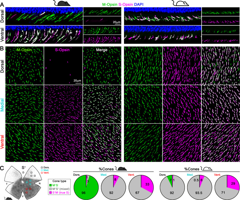
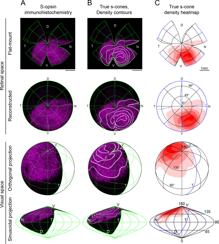
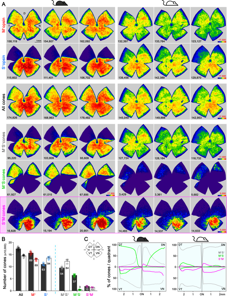
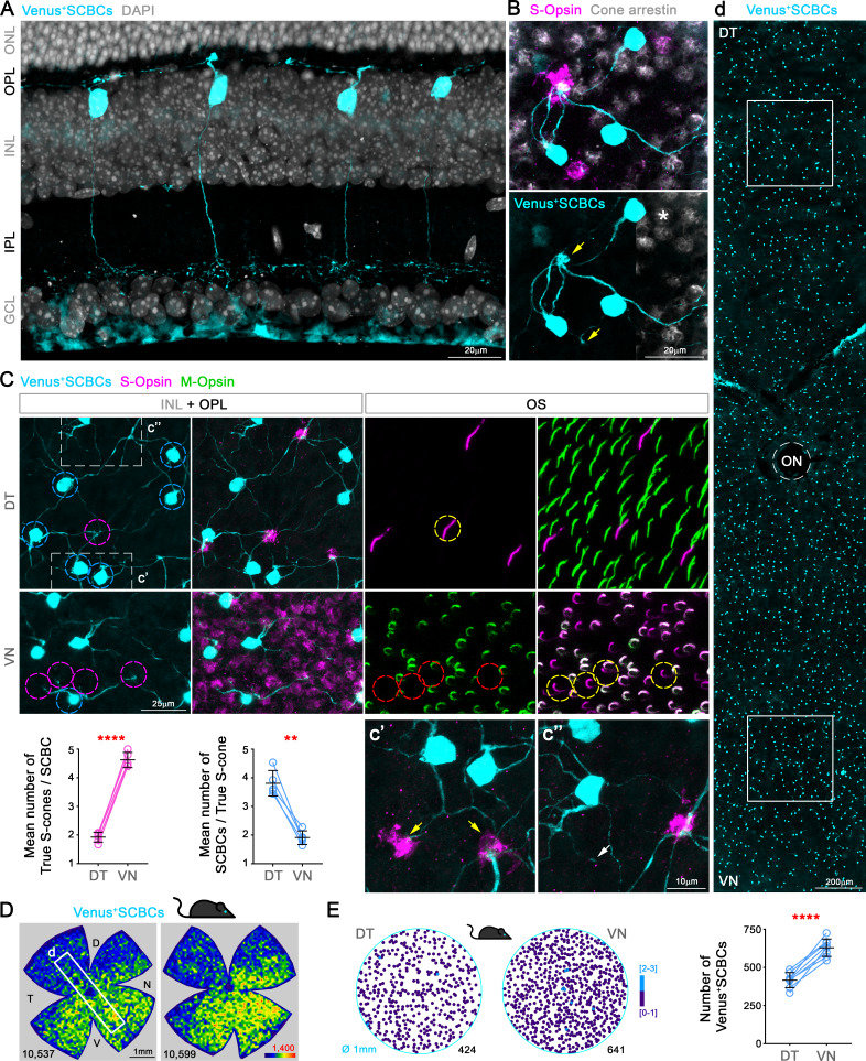
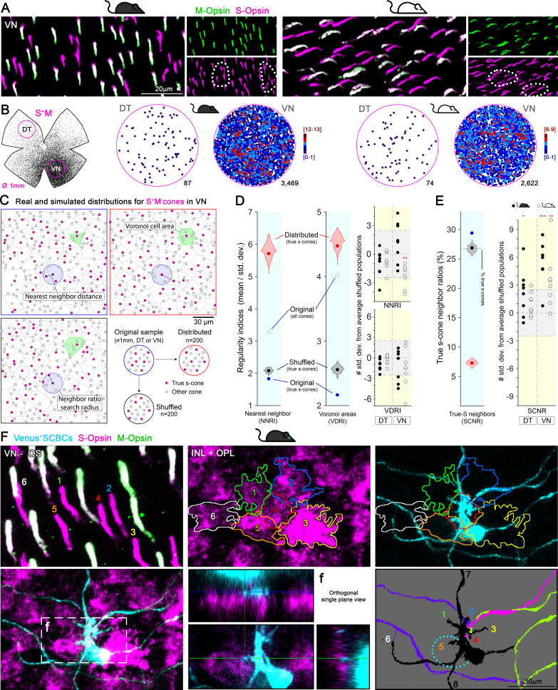
Color, an important visual cue for survival, is encoded by comparing signals from photoreceptors with different spectral sensitivities. The mouse retina expresses a short wavelength-sensitive and a middle/long wavelength-sensitive opsin (S- and M-opsin), forming opposing, overlapping gradients along the dorsal-ventral axis. Here, we analyzed the distribution of all cone types across the entire retina for two commonly used mouse strains. We found, unexpectedly, that 'true S-cones' (S-opsin only) are highly concentrated (up to 30% of cones) in ventral retina. Moreover, S-cone bipolar cells (SCBCs) are also skewed towards ventral retina, with wiring patterns matching the distribution of true S-cones. In addition, true S-cones in the ventral retina form clusters, which may augment synaptic input to SCBCs. Such a unique true S-cone and SCBC connecting pattern forms a basis for mouse color vision, likely reflecting evolutionary adaptation to enhance color coding for the upper visual field suitable for mice's habitat and behavior.

摘要

颜色是生存的重要视觉线索,通过比较具有不同光谱敏感性的光感受器信号来编码。鼠视网膜表达短波长敏感和中/长波长敏感视蛋白(S-和 M-视蛋白),沿背腹轴形成相反的重叠梯度。在这里,我们分析了两种常用小鼠品系整个视网膜中所有视锥细胞类型的分布。出乎意料的是,我们发现“真正的 S 锥细胞”(仅 S-视蛋白)在腹侧视网膜中高度集中(高达 30%的视锥细胞)。此外,S 锥体细胞双极细胞(SCBC)也偏向腹侧视网膜,其布线模式与真正 S 锥细胞的分布相匹配。此外,腹侧视网膜中的真正 S 锥细胞形成簇,这可能会增加对 SCBC 的突触输入。这种独特的真正 S 锥细胞和 SCBC 连接模式为小鼠的颜色视觉形成了基础,可能反映了进化适应,以增强适合小鼠栖息地和行为的上视野的颜色编码。

https://cdn.ncbi.nlm.nih.gov/pmc/blobs/702d/7308094/565249105c10/elife-56840-fig4-figsupp1.jpg
https://cdn.ncbi.nlm.nih.gov/pmc/blobs/702d/7308094/f9375e97ae26/elife-56840-fig1.jpg
https://cdn.ncbi.nlm.nih.gov/pmc/blobs/702d/7308094/d1475c0bcd34/elife-56840-fig2.jpg
https://cdn.ncbi.nlm.nih.gov/pmc/blobs/702d/7308094/4751663d6f2b/elife-56840-fig2-figsupp1.jpg
https://cdn.ncbi.nlm.nih.gov/pmc/blobs/702d/7308094/eb063259b83c/elife-56840-fig3.jpg
https://cdn.ncbi.nlm.nih.gov/pmc/blobs/702d/7308094/8597c457e50d/elife-56840-fig4.jpg
https://cdn.ncbi.nlm.nih.gov/pmc/blobs/702d/7308094/565249105c10/elife-56840-fig4-figsupp1.jpg

相似文献

1
True S-cones are concentrated in the ventral mouse retina and wired for color detection in the upper visual field.真 S 视锥细胞集中在老鼠视网膜的腹侧,并且在上半视野中用于颜色检测。
Elife. 2020 May 28;9:e56840. doi: 10.7554/eLife.56840.
2
The primordial, blue-cone color system of the mouse retina.小鼠视网膜的原始蓝锥细胞颜色系统。
J Neurosci. 2005 Jun 1;25(22):5438-45. doi: 10.1523/JNEUROSCI.1117-05.2005.
3
Retinal cone photoreceptors of the deer mouse Peromyscus maniculatus: development, topography, opsin expression and spectral tuning.鹿鼠 Peromyscus maniculatus 的视网膜圆锥光感受器:发育、拓扑结构、视蛋白表达和光谱调谐。
PLoS One. 2013 Nov 15;8(11):e80910. doi: 10.1371/journal.pone.0080910. eCollection 2013.
4
A mouse M-opsin monochromat: retinal cone photoreceptors have increased M-opsin expression when S-opsin is knocked out.一只小鼠M-视蛋白单色视觉模型:当S-视蛋白被敲除时,视网膜锥状光感受器的M-视蛋白表达增加。
Vision Res. 2011 Feb 23;51(4):447-58. doi: 10.1016/j.visres.2010.12.017. Epub 2011 Jan 8.
5
Maps of cone opsin input to mouse V1 and higher visual areas.视锥蛋白输入到小鼠初级视觉皮层及更高视觉区域的图谱。
J Neurophysiol. 2017 Apr 1;117(4):1674-1682. doi: 10.1152/jn.00849.2016. Epub 2017 Jan 18.
6
Chromatic bipolar cell pathways in the mouse retina.小鼠视网膜中的彩色双极细胞通路。
J Neurosci. 2011 Apr 27;31(17):6504-17. doi: 10.1523/JNEUROSCI.0616-11.2011.
7
Spatial Organization of Chromatic Pathways in the Mouse Dorsal Lateral Geniculate Nucleus.小鼠背外侧膝状核中色觉通路的空间组织
J Neurosci. 2017 Feb 1;37(5):1102-1116. doi: 10.1523/JNEUROSCI.1742-16.2016. Epub 2016 Dec 16.
8
The murine cone photoreceptor: a single cone type expresses both S and M opsins with retinal spatial patterning.小鼠视锥光感受器:单一视锥类型通过视网膜空间模式表达S和M视蛋白。
Neuron. 2000 Sep;27(3):513-23. doi: 10.1016/s0896-6273(00)00062-3.
9
Gene-based Therapy in a Mouse Model of Blue Cone Monochromacy.基于基因的治疗在蓝色锥体单色性的小鼠模型中。
Sci Rep. 2017 Jul 27;7(1):6690. doi: 10.1038/s41598-017-06982-7.
10
Spectral and temporal sensitivity of cone-mediated responses in mouse retinal ganglion cells.小鼠视网膜神经节细胞中视锥介导反应的光谱和时间灵敏度。
J Neurosci. 2011 May 25;31(21):7670-81. doi: 10.1523/JNEUROSCI.0629-11.2011.

引用本文的文献

1
Comparative Analysis of Tyrosine Hydroxylase Amacrine Cells in the Mammalian Retina: Distribution and Quantification in Mouse, Rat, Ground Squirrel and Macaque Retinas.哺乳动物视网膜中酪氨酸羟化酶无长突细胞的比较分析:小鼠、大鼠、地松鼠和猕猴视网膜中的分布与定量
Int J Mol Sci. 2025 Jul 20;26(14):6972. doi: 10.3390/ijms26146972.
2
A Simple and Rapid Method for Simultaneous Isolation of Mouse Retina and RPE Wholemounts.一种同时分离小鼠视网膜和视网膜色素上皮全层标本的简单快速方法。
Transl Vis Sci Technol. 2025 May 1;14(5):14. doi: 10.1167/tvst.14.5.14.
3
A standardized nomenclature for the rods and cones of the vertebrate retina.

本文引用的文献

1
Neural circuits in the mouse retina support color vision in the upper visual field.鼠眼视网膜中的神经回路支持上视野的色觉。
Nat Commun. 2020 Jul 13;11(1):3481. doi: 10.1038/s41467-020-17113-8.
2
Modeling binary and graded cone cell fate patterning in the mouse retina.在小鼠视网膜中对双极细胞和分级锥形细胞命运模式进行建模。
PLoS Comput Biol. 2020 Mar 9;16(3):e1007691. doi: 10.1371/journal.pcbi.1007691. eCollection 2020 Mar.
3
A Color Vision Circuit for Non-Image-Forming Vision in the Primate Retina.灵长类动物视网膜中非成像视觉的色觉电路。
脊椎动物视网膜视杆细胞和视锥细胞的标准化命名法。
PLoS Biol. 2025 May 7;23(5):e3003157. doi: 10.1371/journal.pbio.3003157. eCollection 2025 May.
4
Task-specific regional circuit adaptations in distinct mouse retinal ganglion cells.不同小鼠视网膜神经节细胞中特定任务的区域回路适应性
Sci Adv. 2025 Apr 25;11(17):eadp7075. doi: 10.1126/sciadv.adp7075. Epub 2025 Apr 23.
5
Molecular mechanism establishing the OFF pathway in vision.在视觉中建立OFF通路的分子机制。
Nat Commun. 2025 Apr 18;16(1):3708. doi: 10.1038/s41467-025-59046-0.
6
Long-Term Impairment of Retinal Ganglion Cell Function After Oxygen-Induced Retinopathy.氧诱导性视网膜病变后视网膜神经节细胞功能的长期损害
Cells. 2025 Mar 29;14(7):512. doi: 10.3390/cells14070512.
7
Spatial characterization of RPE structure and lipids in the PEX1-p.Gly844Asp mouse model for Zellweger spectrum disorder.针对齐-韦氏综合征谱障碍的PEX1-p.Gly844Asp小鼠模型中视网膜色素上皮(RPE)结构和脂质的空间特征分析
J Lipid Res. 2025 Apr;66(4):100771. doi: 10.1016/j.jlr.2025.100771. Epub 2025 Mar 7.
8
Spatial distribution and functional integration of displaced retinal ganglion cells.移位视网膜神经节细胞的空间分布与功能整合
Sci Rep. 2025 Feb 28;15(1):7123. doi: 10.1038/s41598-025-91045-5.
9
A Complete Spatial Map of Mouse Retinal Ganglion Cells Reveals Density and Gene Expression Specializations.小鼠视网膜神经节细胞的完整空间图谱揭示了密度和基因表达特化。
bioRxiv. 2025 Feb 12:2025.02.10.637538. doi: 10.1101/2025.02.10.637538.
10
Transcriptional Heterogeneity and Differential Response of Rod Photoreceptor Pathway Uncovered by Single-Cell RNA Sequencing of the Aging Mouse Retina.衰老小鼠视网膜单细胞RNA测序揭示视杆光感受器通路的转录异质性和差异反应
Aging Cell. 2025 May;24(5):e70001. doi: 10.1111/acel.70001. Epub 2025 Feb 15.
Curr Biol. 2020 Apr 6;30(7):1269-1274.e2. doi: 10.1016/j.cub.2020.01.040. Epub 2020 Feb 20.
4
Understanding the retinal basis of vision across species.了解跨物种的视觉视网膜基础。
Nat Rev Neurosci. 2020 Jan;21(1):5-20. doi: 10.1038/s41583-019-0242-1. Epub 2019 Nov 28.
5
An arbitrary-spectrum spatial visual stimulator for vision research.用于视觉研究的任意谱空间视觉刺激器。
Elife. 2019 Sep 23;8:e48779. doi: 10.7554/eLife.48779.
6
Spatial summation of individual cones in human color vision.人类颜色视觉中单锥细胞的空间总和。
PLoS One. 2019 Jul 25;14(7):e0211397. doi: 10.1371/journal.pone.0211397. eCollection 2019.
7
The Retinal Basis of Vertebrate Color Vision.脊椎动物颜色视觉的视网膜基础。
Annu Rev Vis Sci. 2019 Sep 15;5:177-200. doi: 10.1146/annurev-vision-091718-014926. Epub 2019 Jun 21.
8
Zebrafish Differentially Process Color across Visual Space to Match Natural Scenes.斑马鱼在视觉空间中对颜色进行差异化处理以匹配自然场景。
Curr Biol. 2018 Jul 9;28(13):2018-2032.e5. doi: 10.1016/j.cub.2018.04.075. Epub 2018 Jun 21.
9
A novel map of the mouse eye for orienting retinal topography in anatomical space.一种用于在解剖空间中定位视网膜拓扑结构的新型小鼠眼球图谱。
J Comp Neurol. 2018 Aug 1;526(11):1749-1759. doi: 10.1002/cne.24446. Epub 2018 Apr 29.
10
Inhomogeneous Encoding of the Visual Field in the Mouse Retina.在小鼠视网膜中呈现出视野的非均匀编码。
Curr Biol. 2018 Mar 5;28(5):655-665.e3. doi: 10.1016/j.cub.2018.01.016. Epub 2018 Feb 15.