Isildar Basak, Ozkan Serbay, Ercin Merve, Gezginci-Oktayoglu Selda, Oncul Mahmut, Koyuturk Meral
Department of Histology and Embryology, Cerrahpasa Faculty of Medicine, Istanbul University-Cerrahpasa, Istanbul, Turkey.
Department of Biology, Molecular Biology Section, Faculty of Science, Istanbul University, Istanbul, Turkey.
Inflamm Regen. 2022 Nov 30;42(1):55. doi: 10.1186/s41232-022-00241-7.
Type 1 diabetes (T1D) is a T-cell-mediated autoimmune disease characterized by the irreversible destruction of insulin-producing β-cells in pancreatic islets. Helper and cytotoxic T-cells and cytokine production, which is impaired by this process, take a synergetic role in β-cell destruction, and hyperglycemia develops due to insulin deficiency in the body. Mesenchymal stem cells (MSCs) appear like an excellent therapeutic tool for autoimmune diseases with pluripotent, regenerative, and immunosuppressive properties. Paracrine factors released from MSCs play a role in immunomodulation by increasing angiogenesis and proliferation and suppressing apoptosis. In this context, the study aims to investigate the therapeutic effects of MSC's secretomes by conditioned medium (CM) obtained from human umbilical cord-derived MSCs cultured in 2-dimensional (2D) and 3-dimensional (3D) environments in the T1D model.
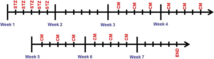
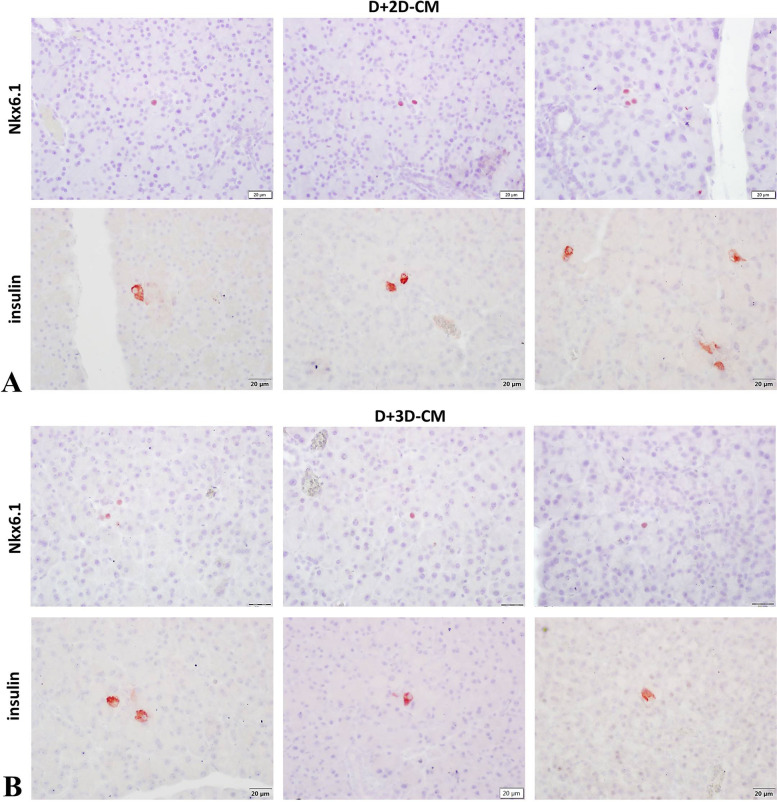
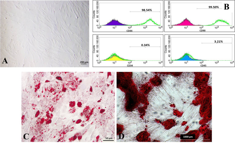
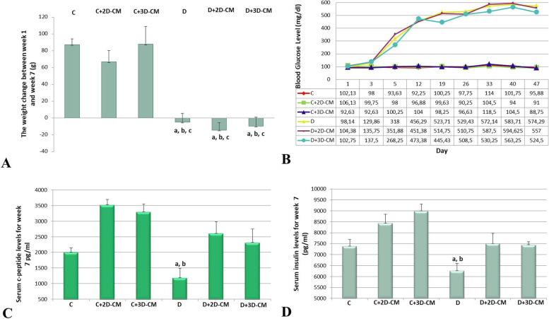
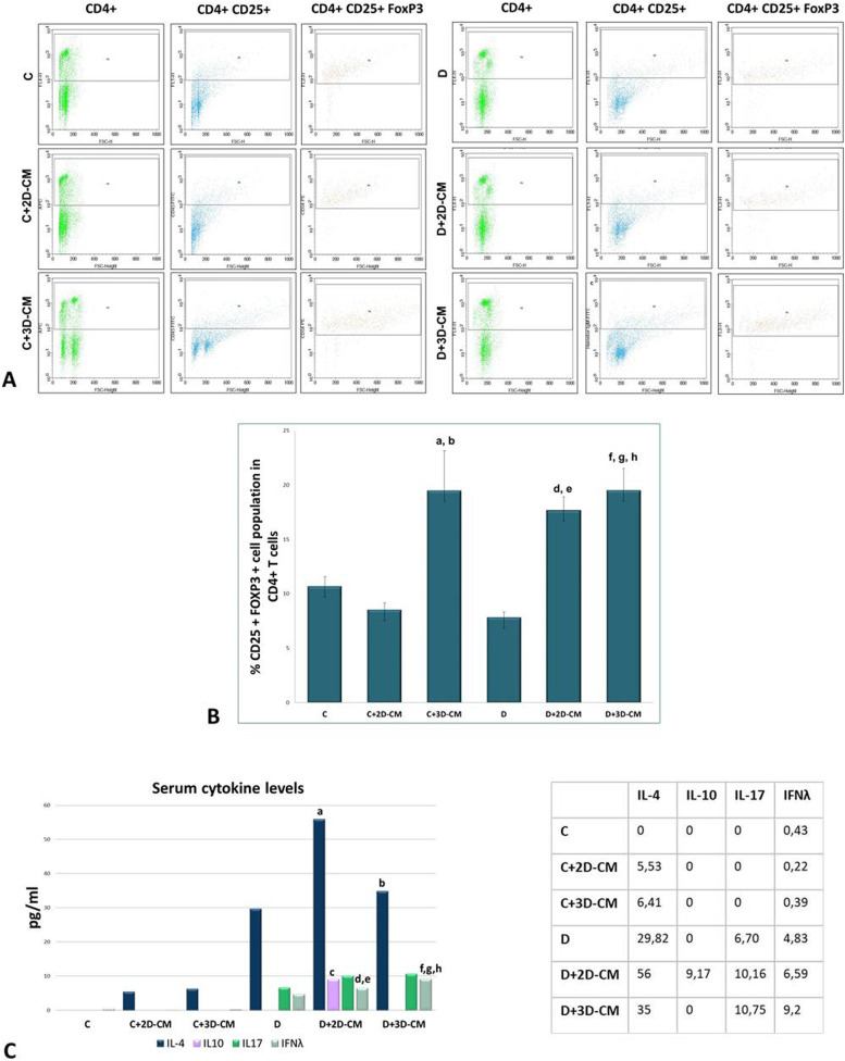
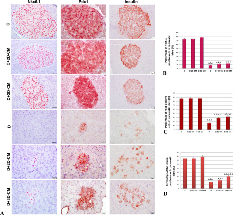
First, MSCs were isolated from the human umbilical cord, and the cells were characterized. Then, two different CMs were prepared by culturing MSCs in 2D and 3D environments. The CM contents were analyzed in terms of total protein, IL-4, IL-10, IL-17, and IFN-λ. In vivo studies were performed in Sprague-Dawley-type rats with an autoimmune T1D model, and twelve doses of CM were administered intraperitoneally for 4 weeks within the framework of a particular treatment model. In order to evaluate immunomodulation, the Treg population was determined in lymphocytes isolated from the spleen after sacrification, and IL-4, IL-10, IL-17, and IFN-λ cytokines were analyzed in serum. Finally, β-cell regeneration was evaluated immunohistochemically by labeling Pdx1, Nkx6.1, and insulin markers, which are critical for the formation of β-cells.
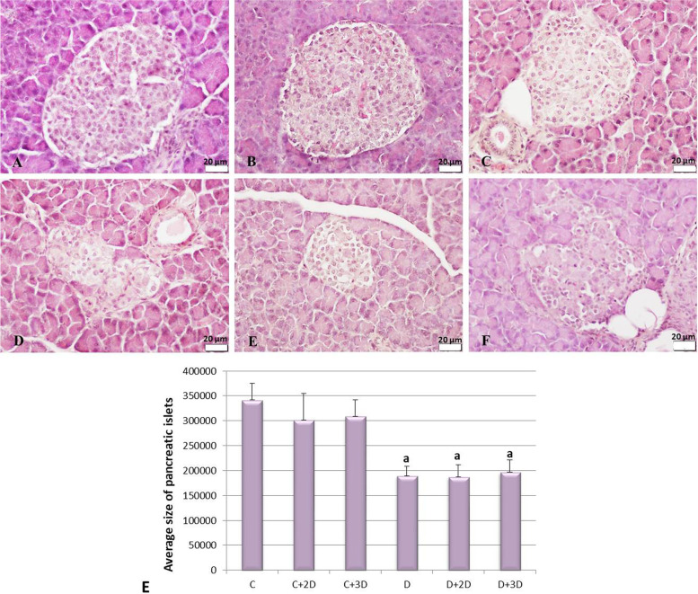
Total protein and IL-4 levels were higher in 3D-CM compared to 2D-CM. In vivo results showed that CMs induce the Treg population and regulate cytokine release. When the immunohistochemical results were evaluated together, it was determined that CM application significantly increased the rate of β-cells in the islets. This increase was at the highest level in the 3D-CM applied group.
The dual therapeutic effect of MSC-CM on immunomodulation and homeostasis/regeneration of β-cells in the T1D model has been demonstrated. Furthermore, this effect could be improved by using 3D scaffolds for culturing MSCs while preparing CM.
1型糖尿病(T1D)是一种由T细胞介导的自身免疫性疾病,其特征是胰岛中产生胰岛素的β细胞发生不可逆破坏。辅助性T细胞和细胞毒性T细胞以及在此过程中受损的细胞因子产生,在β细胞破坏中起协同作用,并且由于体内胰岛素缺乏而导致高血糖症。间充质干细胞(MSCs)似乎是一种具有多能性、再生性和免疫抑制特性的自身免疫性疾病的优秀治疗工具。间充质干细胞释放的旁分泌因子通过增加血管生成和增殖以及抑制细胞凋亡在免疫调节中发挥作用。在此背景下,本研究旨在探讨在T1D模型中,从二维(2D)和三维(3D)环境中培养的人脐带源间充质干细胞获得的条件培养基(CM)对间充质干细胞分泌组的治疗效果。
首先,从人脐带中分离间充质干细胞,并对细胞进行表征。然后,通过在2D和3D环境中培养间充质干细胞制备两种不同的CM。从总蛋白、白细胞介素-4(IL-4)、白细胞介素-10(IL-10)、白细胞介素-17(IL-17)和干扰素-λ(IFN-λ)方面分析CM的含量。在具有自身免疫性T1D模型的Sprague-Dawley型大鼠中进行体内研究,并在特定治疗模型的框架内腹腔注射12剂CM,持续4周。为了评估免疫调节,在处死动物后从脾脏分离的淋巴细胞中测定调节性T细胞(Treg)群体,并分析血清中的IL-4、IL-10、IL-17和IFN-λ细胞因子。最后,通过标记对β细胞形成至关重要的胰腺十二指肠同源盒1(Pdx1)、NK6转录因子相关蛋白1(Nkx6.1)和胰岛素标志物,免疫组织化学评估β细胞再生。
与2D-CM相比,3D-CM中的总蛋白和IL-4水平更高。体内结果表明,CM可诱导Treg群体并调节细胞因子释放。综合评估免疫组织化学结果时,确定CM应用显著增加了胰岛中β细胞的比例。在应用3D-CM的组中,这种增加处于最高水平。
已证明间充质干细胞条件培养基对T1D模型中β细胞的免疫调节和内环境稳定/再生具有双重治疗作用。此外,在制备CM时使用3D支架培养间充质干细胞可以提高这种效果。