• 文献检索
  • 文档翻译
  • 深度研究
  • 学术资讯
  • Suppr Zotero 插件Zotero 插件
  • 邀请有礼
  • 套餐&价格
  • 历史记录
应用&插件
Suppr Zotero 插件Zotero 插件浏览器插件Mac 客户端Windows 客户端微信小程序
定价
高级版会员购买积分包购买API积分包
服务
文献检索文档翻译深度研究API 文档MCP 服务
关于我们
关于 Suppr公司介绍联系我们用户协议隐私条款
关注我们

Suppr 超能文献

核心技术专利:CN118964589B侵权必究
粤ICP备2023148730 号-1Suppr @ 2026

文献检索

告别复杂PubMed语法,用中文像聊天一样搜索,搜遍4000万医学文献。AI智能推荐,让科研检索更轻松。

立即免费搜索

文件翻译

保留排版,准确专业,支持PDF/Word/PPT等文件格式,支持 12+语言互译。

免费翻译文档

深度研究

AI帮你快速写综述,25分钟生成高质量综述,智能提取关键信息,辅助科研写作。

立即免费体验

基于深度学习的翼状胬肉筛查与病变区域分割。

Pterygium Screening and Lesion Area Segmentation Based on Deep Learning.

机构信息

School of Information Engineering, Huzhou University, Huzhou 313000, China.

Zhejiang Province Key Laboratory of Smart Management & Application of Modern Agricultural Resources, Huzhou University, Huzhou 313000, China.

出版信息

J Healthc Eng. 2022 Nov 21;2022:3942110. doi: 10.1155/2022/3942110. eCollection 2022.

DOI:10.1155/2022/3942110
PMID:36451763
原文链接:https://pmc.ncbi.nlm.nih.gov/articles/PMC9705081/
Abstract

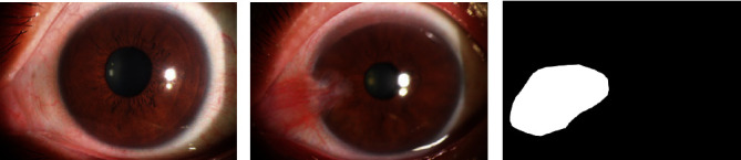
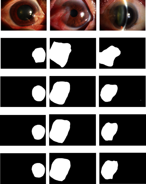
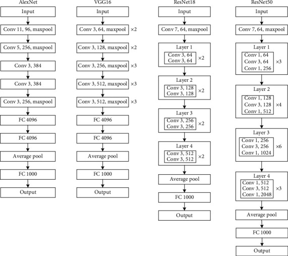
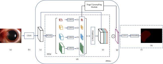
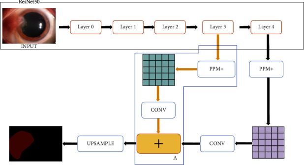
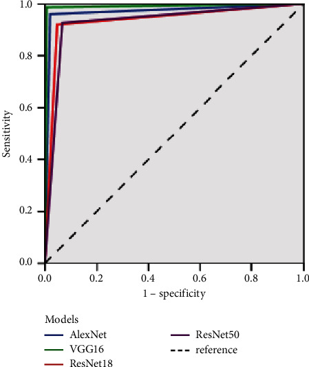
A two-category model and a segmentation model of pterygium were proposed to assist ophthalmologists in establishing the diagnosis of ophthalmic diseases. A total of 367 normal anterior segment images and 367 pterygium anterior segment images were collected at the Affiliated Eye Hospital of Nanjing Medical University. AlexNet, VGG16, ResNet18, and ResNet50 models were used to train the two-category pterygium models. A total of 150 normal and 150 pterygium anterior segment images were used to test the models, and the results were compared. The main evaluation indicators, including sensitivity, specificity, area under the curve, kappa value, and receiver operator characteristic curves of the four models, were compared. Simultaneously, 367 pterygium anterior segment images were used to train two improved pterygium segmentation models based on PSPNet. A total of 150 pterygium images were used to test the models, and the results were compared with those of the other four segmentation models. The main evaluation indicators included mean intersection over union (MIOU), IOU, mean average precision (MPA), and PA. Among the two-category models of pterygium, the best diagnostic result was obtained using the VGG16 model. The diagnostic accuracy, kappa value, diagnostic sensitivity of pterygium, diagnostic specificity of pterygium, and F1-score were 99%, 98%, 98.67%, 99.33%, and 99%, respectively. Among the pterygium segmentation models, the double phase-fusion PSPNet model had the best results, with MIOU, IOU, MPA, and PA of 86.57%, 78.1%, 92.3%, and 86.96%, respectively. This study designed a pterygium two-category model and a pterygium segmentation model for the images of the normal anterior and pterygium anterior segments, which could help patients self-screen easily and assist ophthalmologists in establishing the diagnosis of ophthalmic diseases and marking the actual scope of surgery.

摘要

提出了一种翼状胬肉的二分类模型和分割模型,以协助眼科医生建立眼病的诊断。在南京医科大学附属医院收集了 367 张正常眼前节图像和 367 张翼状胬肉眼前节图像。使用 AlexNet、VGG16、ResNet18 和 ResNet50 模型训练二分类翼状胬肉模型。使用 150 张正常和 150 张翼状胬肉眼前节图像测试模型,并比较结果。比较了四个模型的主要评估指标,包括敏感性、特异性、曲线下面积、kappa 值和受试者工作特征曲线。同时,使用 367 张翼状胬肉眼前节图像基于 PSPNet 训练两个改进的翼状胬肉分割模型。使用 150 张翼状胬肉图像测试模型,并与其他四个分割模型的结果进行比较。主要评估指标包括平均交并比(MIOU)、IOU、平均准确率(MPA)和 PA。在翼状胬肉的二分类模型中,VGG16 模型获得了最佳诊断结果。翼状胬肉的诊断准确率、kappa 值、诊断敏感性、诊断特异性和 F1 评分分别为 99%、98%、98.67%、99.33%和 99%。在翼状胬肉分割模型中,双相融合 PSPNet 模型效果最好,MIOU、IOU、MPA 和 PA 分别为 86.57%、78.1%、92.3%和 86.96%。本研究为正常眼前节和翼状胬肉眼前节图像设计了翼状胬肉二分类模型和分割模型,方便患者进行自我筛查,并协助眼科医生建立眼病的诊断,标记实际手术范围。

https://cdn.ncbi.nlm.nih.gov/pmc/blobs/f282/9705081/0de2f5881ca8/JHE2022-3942110.006.jpg
https://cdn.ncbi.nlm.nih.gov/pmc/blobs/f282/9705081/082d50364a35/JHE2022-3942110.001.jpg
https://cdn.ncbi.nlm.nih.gov/pmc/blobs/f282/9705081/b878c7af7f7a/JHE2022-3942110.002.jpg
https://cdn.ncbi.nlm.nih.gov/pmc/blobs/f282/9705081/c7711bcfc47b/JHE2022-3942110.003.jpg
https://cdn.ncbi.nlm.nih.gov/pmc/blobs/f282/9705081/6d2a484fc794/JHE2022-3942110.004.jpg
https://cdn.ncbi.nlm.nih.gov/pmc/blobs/f282/9705081/53d2e8daa10b/JHE2022-3942110.005.jpg
https://cdn.ncbi.nlm.nih.gov/pmc/blobs/f282/9705081/0de2f5881ca8/JHE2022-3942110.006.jpg

相似文献

1
Pterygium Screening and Lesion Area Segmentation Based on Deep Learning.基于深度学习的翼状胬肉筛查与病变区域分割。
J Healthc Eng. 2022 Nov 21;2022:3942110. doi: 10.1155/2022/3942110. eCollection 2022.
2
Research on an Intelligent Lightweight-Assisted Pterygium Diagnosis Model Based on Anterior Segment Images.基于眼前段图像的智能轻量化翼状胬肉诊断模型研究。
Dis Markers. 2021 Jul 29;2021:7651462. doi: 10.1155/2021/7651462. eCollection 2021.
3
Intelligent diagnostic model for pterygium by combining attention mechanism and MobileNetV2.基于注意力机制与MobileNetV2相结合的翼状胬肉智能诊断模型
Int J Ophthalmol. 2024 Jul 18;17(7):1184-1192. doi: 10.18240/ijo.2024.07.02. eCollection 2024.
4
Implementation and Application of an Intelligent Pterygium Diagnosis System Based on Deep Learning.基于深度学习的智能翼状胬肉诊断系统的实现与应用
Front Psychol. 2021 Oct 22;12:759229. doi: 10.3389/fpsyg.2021.759229. eCollection 2021.
5
A Novel System for Measuring Pterygium's Progress Using Deep Learning.一种使用深度学习测量翼状胬肉进展的新型系统。
Front Med (Lausanne). 2022 Feb 14;9:819971. doi: 10.3389/fmed.2022.819971. eCollection 2022.
6
Automated pterygium detection method of anterior segment photographed images.眼前节拍摄图像的翼状胬肉自动检测方法。
Comput Methods Programs Biomed. 2018 Feb;154:71-78. doi: 10.1016/j.cmpb.2017.10.026. Epub 2017 Oct 31.
7
Screening of Common Retinal Diseases Using Six-Category Models Based on EfficientNet.基于EfficientNet的六类模型用于常见视网膜疾病的筛查
Front Med (Lausanne). 2022 Feb 23;9:808402. doi: 10.3389/fmed.2022.808402. eCollection 2022.
8
A deep learning-based model for automatic segmentation and evaluation of corneal neovascularization using slit-lamp anterior segment images.一种基于深度学习的模型,用于使用裂隙灯眼前节图像对角膜新生血管进行自动分割和评估。
Quant Imaging Med Surg. 2023 Oct 1;13(10):6778-6788. doi: 10.21037/qims-23-99. Epub 2023 Aug 25.
9
Application of artificial intelligence models for detecting the pterygium that requires surgical treatment based on anterior segment images.基于眼前节图像的人工智能模型在检测需要手术治疗的翼状胬肉中的应用。
Front Neurosci. 2022 Dec 20;16:1084118. doi: 10.3389/fnins.2022.1084118. eCollection 2022.
10
A comparative study of pre-trained convolutional neural networks for semantic segmentation of breast tumors in ultrasound.用于超声乳腺肿瘤语义分割的预训练卷积神经网络的比较研究
Comput Biol Med. 2020 Nov;126:104036. doi: 10.1016/j.compbiomed.2020.104036. Epub 2020 Oct 8.

引用本文的文献

1
Clinical Applications of Artificial Intelligence in Corneal Diseases.人工智能在角膜疾病中的临床应用
Vision (Basel). 2025 Aug 18;9(3):71. doi: 10.3390/vision9030071.
2
A semantic segmentation-based automatic pterygium assessment and grading system.一种基于语义分割的翼状胬肉自动评估与分级系统。
Front Med (Lausanne). 2025 Mar 13;12:1507226. doi: 10.3389/fmed.2025.1507226. eCollection 2025.
3
A Performance Evaluation of Convolutional Neural Network Architectures for Pterygium Detection in Anterior Segment Eye Images.用于眼前节图像中翼状胬肉检测的卷积神经网络架构性能评估

本文引用的文献

1
EyeHealer: A large-scale anterior eye segment dataset with eye structure and lesion annotations.EyeHealer:一个具有眼部结构和病变注释的大规模眼前节数据集。
Precis Clin Med. 2021 Apr 27;4(2):85-92. doi: 10.1093/pcmedi/pbab009. eCollection 2021 Jun.
2
Joint optic disk and cup segmentation for glaucoma screening using a region-based deep learning network.基于区域的深度学习网络在青光眼筛查中的联合视盘和杯分割。
Eye (Lond). 2023 Apr;37(6):1080-1087. doi: 10.1038/s41433-022-02055-w. Epub 2022 Apr 18.
3
Simultaneous segmentation and classification of the retinal arteries and veins from color fundus images.
Diagnostics (Basel). 2024 Sep 13;14(18):2026. doi: 10.3390/diagnostics14182026.
4
A deep learning-powered diagnostic model for acute pancreatitis.基于深度学习的急性胰腺炎诊断模型。
BMC Med Imaging. 2024 Jun 20;24(1):154. doi: 10.1186/s12880-024-01339-9.
5
Application and visualization study of an intelligence-assisted classification model for common eye diseases using B-mode ultrasound images.基于B超图像的常见眼科疾病智能辅助分类模型的应用与可视化研究
Front Neurosci. 2024 May 14;18:1339075. doi: 10.3389/fnins.2024.1339075. eCollection 2024.
6
Potential applications of artificial intelligence in image analysis in cornea diseases: a review.人工智能在角膜疾病图像分析中的潜在应用:综述
Eye Vis (Lond). 2024 Mar 7;11(1):10. doi: 10.1186/s40662-024-00376-3.
7
Artificial intelligence assisted pterygium diagnosis: current status and perspectives.人工智能辅助翼状胬肉诊断:现状与展望
Int J Ophthalmol. 2023 Sep 18;16(9):1386-1394. doi: 10.18240/ijo.2023.09.04. eCollection 2023.
8
Automated measurement of the disc-fovea angle based on DeepLabv3.基于深度卷积神经网络(DeepLabv3)的视盘-黄斑中心凹夹角自动测量
Front Neurol. 2022 Jul 27;13:949805. doi: 10.3389/fneur.2022.949805. eCollection 2022.
彩色眼底图像中视网膜动脉和静脉的同时分割和分类。
Artif Intell Med. 2021 Aug;118:102116. doi: 10.1016/j.artmed.2021.102116. Epub 2021 May 29.
4
Automatic detection of 39 fundus diseases and conditions in retinal photographs using deep neural networks.使用深度神经网络自动检测视网膜照片中的 39 种眼底疾病和病变。
Nat Commun. 2021 Aug 10;12(1):4828. doi: 10.1038/s41467-021-25138-w.
5
Research on an Intelligent Lightweight-Assisted Pterygium Diagnosis Model Based on Anterior Segment Images.基于眼前段图像的智能轻量化翼状胬肉诊断模型研究。
Dis Markers. 2021 Jul 29;2021:7651462. doi: 10.1155/2021/7651462. eCollection 2021.
6
Group and Shuffle Convolutional Neural Networks with Pyramid Pooling Module for Automated Pterygium Segmentation.基于金字塔池化模块的分组与随机卷积神经网络用于翼状胬肉自动分割
Diagnostics (Basel). 2021 Jun 17;11(6):1104. doi: 10.3390/diagnostics11061104.
7
Bibliometric analysis and mapping knowledge domain of pterygium: 2000-2019.翼状胬肉的文献计量分析与知识图谱构建:2000 - 2019年
Int J Ophthalmol. 2021 Jun 18;14(6):903-914. doi: 10.18240/ijo.2021.06.17. eCollection 2021.
8
Five-Category Intelligent Auxiliary Diagnosis Model of Common Fundus Diseases Based on Fundus Images.基于眼底图像的常见眼底疾病五类智能辅助诊断模型。
Transl Vis Sci Technol. 2021 Jun 1;10(7):20. doi: 10.1167/tvst.10.7.20.
9
Automated segmentation of retinal nonperfusion area in fluorescein angiography in retinal vein occlusion using convolutional neural networks.利用卷积神经网络对视网膜静脉阻塞荧光素血管造影中的视网膜无灌注区进行自动分割。
Med Phys. 2021 Feb;48(2):648-658. doi: 10.1002/mp.14640. Epub 2020 Dec 23.
10
An Evaluation System of Fundus Photograph-Based Intelligent Diagnostic Technology for Diabetic Retinopathy and Applicability for Research.基于眼底照片的糖尿病视网膜病变智能诊断技术评估系统及其研究适用性
Diabetes Ther. 2019 Oct;10(5):1811-1822. doi: 10.1007/s13300-019-0652-0. Epub 2019 Jul 9.