University of South Florida, Department of Dermatology and Cutaneous Surgery, Tampa, Florida, United States.
Moffitt Cancer Center, Cutaneous Oncology, Tampa, Florida, United States.
J Biomed Opt. 2022 Aug;27(8):080902. doi: 10.1117/1.JBO.27.8.080902. Epub 2022 Aug 8.
Dermoscopes incorporate light, polarizers, and optical magnification into a handheld tool that is commonly used by dermatologists to evaluate skin findings. Diagnostic accuracy is improved when dermoscopes are used, and some major artificial intelligence (AI) projects have been accomplished using dermocopic images. Color rendering consistency and fidelity are crucial for clinical diagnostics, AI, and image processing applications.
With many devices available on the market, our objective was to measure the emission spectra of various dermoscopes, compare them with other light sources, and illustrate variations in reflected colors from images of a reference sample.
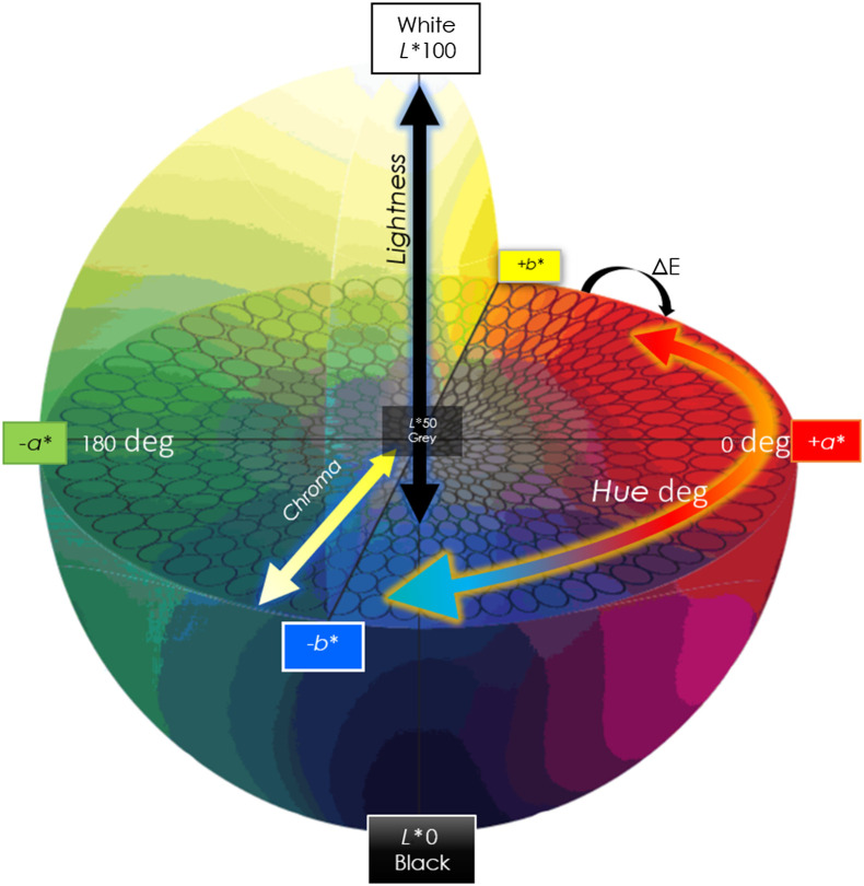
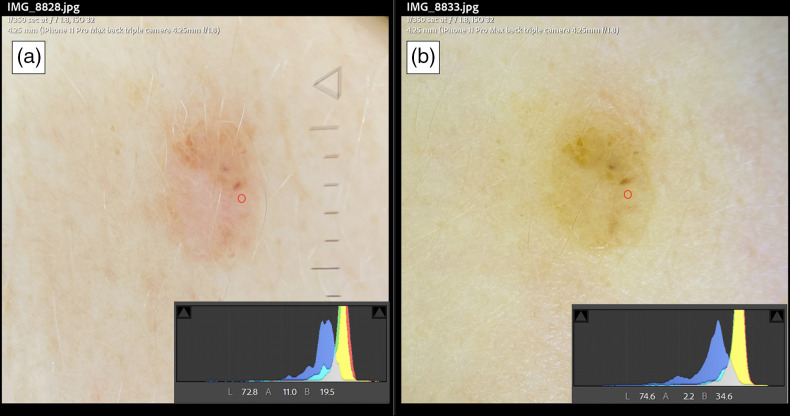
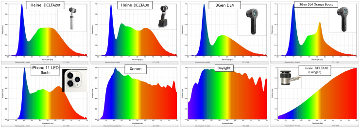
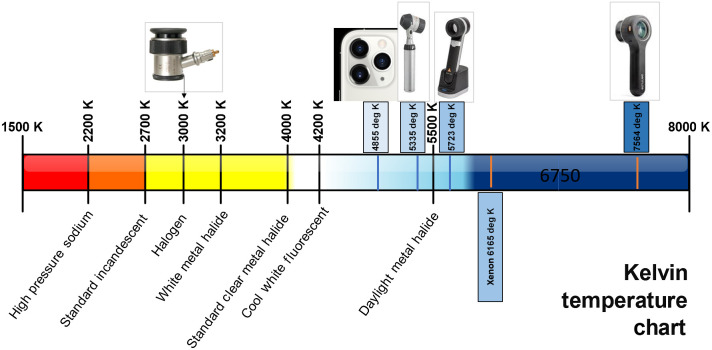
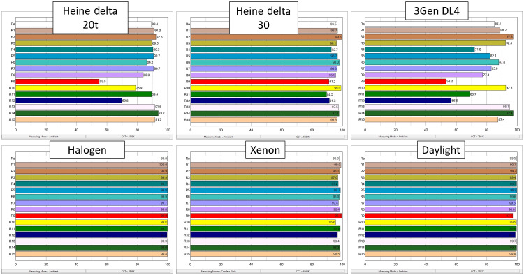
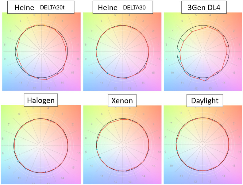
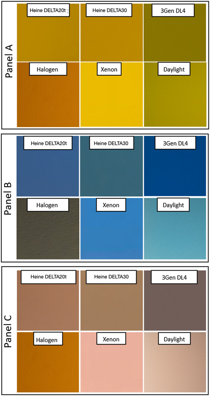
A spectrometer measured the spectral power distribution (SPD) produced by four dermoscope models and three alternate light sources, illustrating differences in the emission spectra. Most dermoscopes use light-emitting diodes (LEDs), which are inconsistent when compared with one another. An LED was compared with halogen, xenon-arc, and daylight sources. Images of a micro ColorChecker were acquired from several sources, and three specific colors were selected to compare in CIELAB color space. Color consistency and color fidelity measured by color rendering index (CRI) and TM-30-18 graphical vectors show variation in saturation and chroma fidelity.
A marked degree of variation was observed in both the emission and reflected light coming from different dermoscopes and compared with other sources. The same chromophores appeared differently depending on the light source used.
A lack of uniform illumination resulted in inconsistent image color and likely impacted metamerism and visibility of skin chromophores in real-world settings. Artificial light in skin examinations, especially LEDs, may present challenges for the visual separation of specific colors. Attention to LEDs SPD may be important, especially as the field increases dependency on machine/computer vision.
共聚焦显微镜将光、偏振器和光学放大集成到一个手持工具中,皮肤科医生通常用它来评估皮肤发现。使用共聚焦显微镜可以提高诊断准确性,一些主要的人工智能 (AI) 项目已经使用共聚焦显微镜图像完成。颜色渲染的一致性和保真度对于临床诊断、人工智能和图像处理应用至关重要。
市面上有许多设备,我们的目标是测量各种共聚焦显微镜的发射光谱,将其与其他光源进行比较,并说明参考样本图像反射颜色的变化。
光谱仪测量了四个共聚焦显微镜模型和三个替代光源产生的光谱功率分布 (SPD),说明了发射光谱的差异。大多数共聚焦显微镜使用发光二极管 (LED),彼此之间不一致。将 LED 与卤素、氙弧和日光光源进行了比较。从多个来源获取了微 ColorChecker 的图像,并选择了三个特定颜色在 CIELAB 颜色空间中进行比较。由显色指数 (CRI) 和 TM-30-18 图形向量测量的颜色一致性和颜色保真度显示了饱和度和色度保真度的变化。
从不同的共聚焦显微镜以及与其他光源的比较中观察到发射光和反射光都有明显的变化。同一色素因所用光源而异。
照明不均匀导致图像颜色不一致,可能会影响实际环境中皮肤色素的同色异谱和可见度。皮肤检查中的人工光,尤其是 LED,可能会对特定颜色的视觉分离提出挑战。对 LED SPD 的关注可能很重要,尤其是在该领域越来越依赖机器/计算机视觉的情况下。