Department of Visceral, Transplant, Thoracic and Vascular Surgery, University Hospital of Leipzig, Leipzig, Germany.
Department of Orthopedics, Trauma and Plastic Surgery, University Hospital Leipzig, Leipzig, Germany.
PLoS One. 2022 Dec 1;17(12):e0276978. doi: 10.1371/journal.pone.0276978. eCollection 2022.
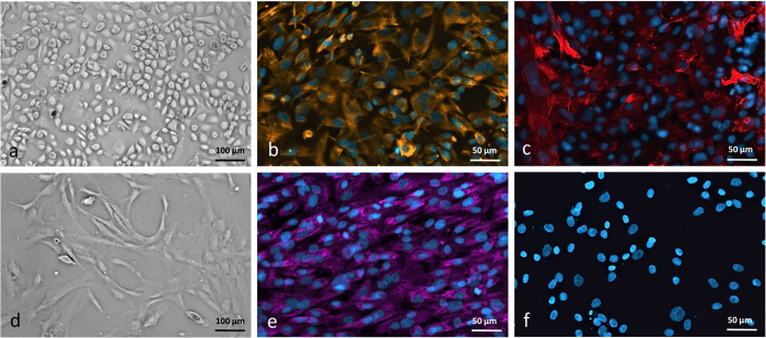
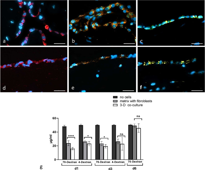
Pleural mesothelial cells are the predominant cell type in the pleural cavity, but their role in the pathogenesis of pleural diseases needs to be further elucidated. 3D organotypic models are an encouraging approach for an in vivo understanding of molecular disease development. The aim of the present study was to develop a 3D organotypic model of the pleural mesothelium. Specimens of human pleura parietalis were obtained from patients undergoing surgery at the University Hospital Leipzig, Germany. 3D co-culture model of pleura was established from human pleural mesothelial cells and fibroblasts. The model was compared to human pleura tissue by phase-contrast and light microscopy, immunochemistry and -fluorescence as well as solute permeation test. Histological assessment of the 3D co-culture model displayed the presence of both cell types mimicking the morphology of the human pleura. Vimentin and Cytokeratin, PHD1 showed a similar expression pattern in pleural biopsies and 3D model. Expression of Ki-67 indicates the presence of proliferating cells. Tight junctional marker ZO-1 was found localized at contact zones between mesothelial cells. Each of these markers were expressed in both the 3D co-culture model and human biopsies. Permeability of 3D organotypic co-culture model of pleura was found to be higher for 70 kDa-Dextran and no significant difference was seen in the permeability for small dextran (4 kDa). In summary, the presented 3D organoid of pleura functions as a robust assay for pleural research serving as a precise reproduction of the in vivo morphology and microenvironment.
胸膜间皮细胞是胸膜腔中的主要细胞类型,但它们在胸膜疾病发病机制中的作用仍需进一步阐明。3D 器官型模型是研究分子疾病发展的体内理解的一种有前途的方法。本研究旨在开发胸膜间皮的 3D 器官型模型。从德国莱比锡大学医院接受手术的患者中获得人胸膜壁层的标本。从人胸膜间皮细胞和成纤维细胞建立了胸膜的 3D 共培养模型。通过相差和明场显微镜、免疫化学和荧光以及溶质渗透试验将该模型与人体胸膜组织进行比较。3D 共培养模型的组织学评估显示存在两种细胞类型,模拟了人体胸膜的形态。波形蛋白和细胞角蛋白、PHD1 在胸膜活检和 3D 模型中表现出相似的表达模式。Ki-67 的表达表明存在增殖细胞。紧密连接标记物 ZO-1 位于间皮细胞之间的接触区。这些标志物均在 3D 共培养模型和人体活检中表达。发现胸膜的 3D 器官型共培养模型对 70 kDa-葡聚糖的通透性较高,而对小葡聚糖(4 kDa)的通透性没有显著差异。总之,所提出的胸膜 3D 类器官可作为胸膜研究的有力检测手段,可精确再现体内形态和微环境。