Division of Respiratory Medicine, Juntendo University Faculty of Medicine and Graduate School of Medicine, 3-1-3 Hongo, Bunkyo-ku, Tokyo, 113-8431, Japan.
The Study Group for Pneumothorax and Cystic Lung Diseases, Tokyo, 158-0095, Japan.
Sci Rep. 2021 May 24;11(1):10814. doi: 10.1038/s41598-021-90184-9.
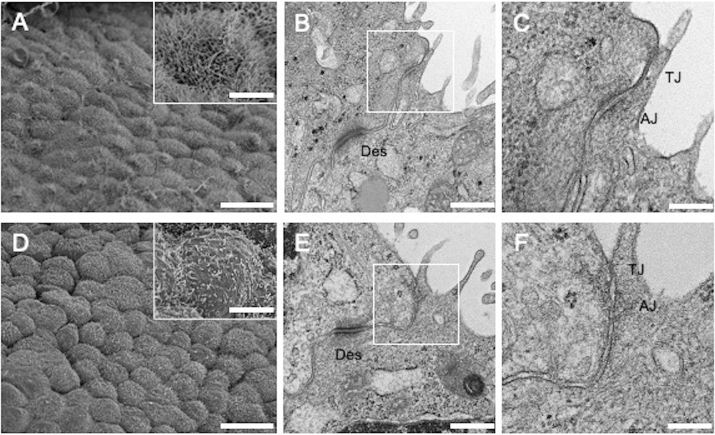
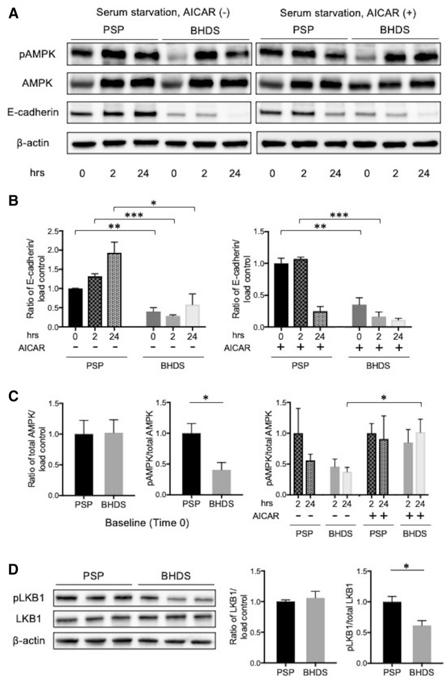
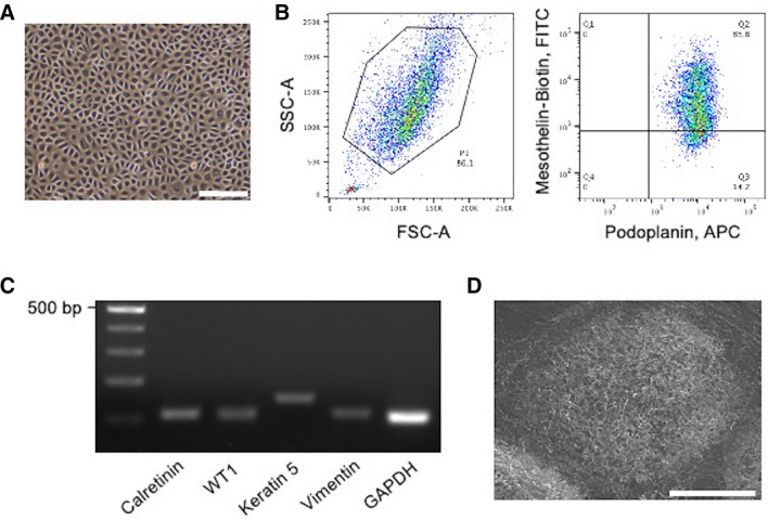
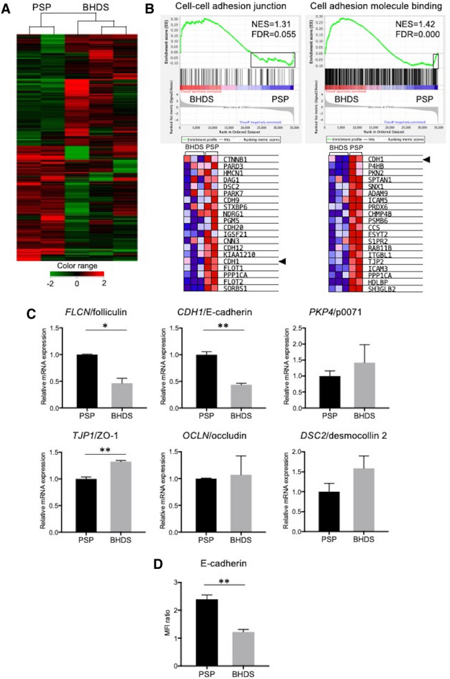
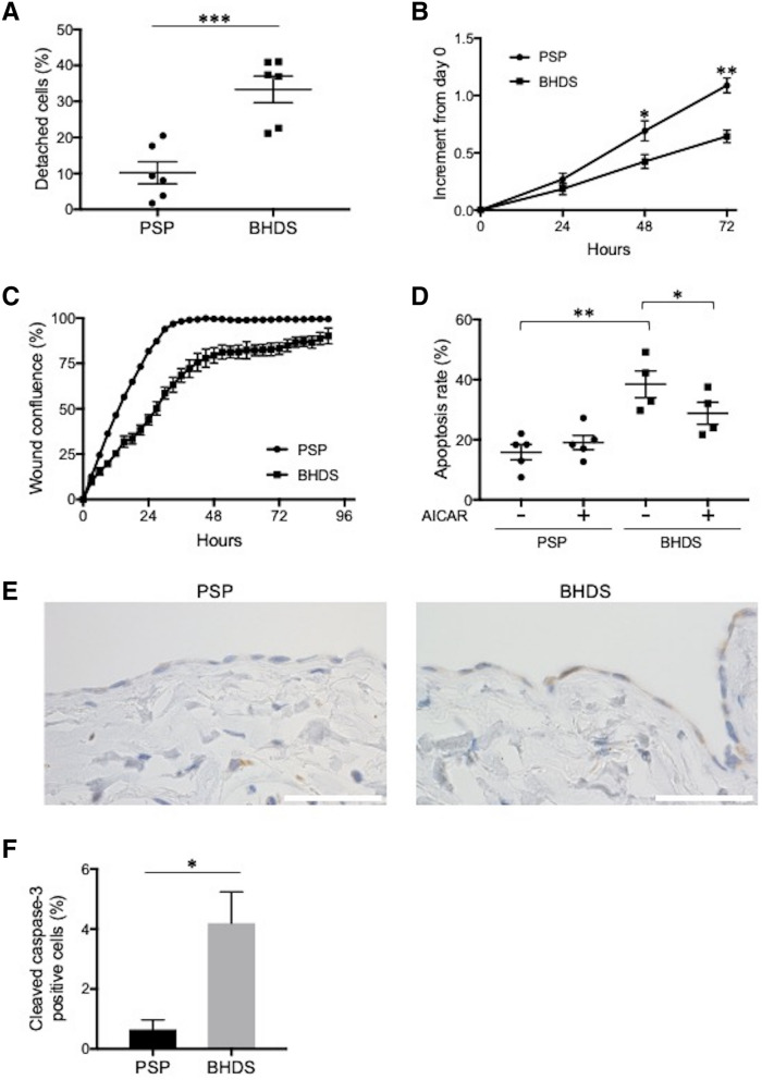
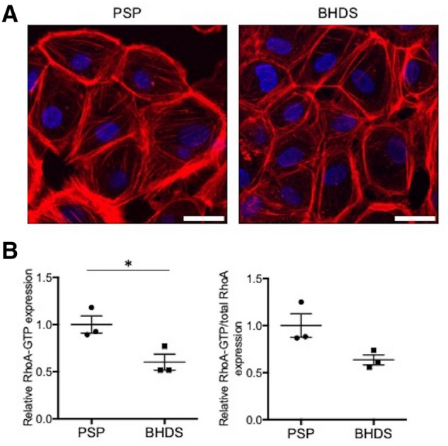
Birt-Hogg-Dubé syndrome (BHDS), an autosomal dominant inheritance disease caused by folliculin (FLCN) mutations, is associated with lung cysts and spontaneous pneumothorax. The possibility of FLCN haploinsufficiency in pleural mesothelial cells (PMCs) contributing to development of pneumothorax has not yet been clarified. Electron microscopy revealed exposed intercellular boundaries between PMCs on visceral pleura and decreased electron density around the adherens junctions in BHDS. To characterize cellular function of PMCs in BHDS patients (BHDS-PMCs), during surgery for pneumothorax, we established the flow cytometry-based methods of isolating high-purity PMCs from pleural lavage fluid. BHDS-PMCs showed impaired cell attachment and a significant decrease in proliferation and migration, but a significant increase in apoptosis compared with PMCs from primary spontaneous pneumothorax (PSP) patients (PSP-PMCs). Microarray analysis using isolated PMCs revealed a significant alteration in the expression of genes belonging to Gene Ontology terms "cell-cell adhesion junction" and "cell adhesion molecule binding". Gene set enrichment analysis demonstrated that CDH1, encoding E-cadherin, was identified in the down-regulated leading edge of a plot in BHDS-PMCs. AMPK and LKB1 activation were significantly impaired in BHDS-PMCs compared with PSP-PMCs. Our findings indicate that FLCN haploinsufficiency may affect the E-cadherin-LKB1-AMPK axis and lead to abnormal cellular function in BHDS-PMCs.
Birt-Hogg-Dubé 综合征(BHDS)是一种常染色体显性遗传疾病,由滤泡素(FLCN)突变引起,与肺囊肿和自发性气胸有关。FLCN 部分功能不足是否会导致胸膜间皮细胞(PMCs)异常,从而导致气胸的发生,目前尚不清楚。电子显微镜显示 BHDS 的脏层胸膜 PMCs 细胞间边界暴露,黏着连接周围的电子密度降低。为了研究 BHDS 患者(BHDS-PMCs)的 PMCs 细胞功能,我们在气胸手术期间,建立了基于流式细胞术从胸腔灌洗液中分离高纯度 PMCs 的方法。与原发性自发性气胸(PSP)患者(PSP-PMCs)相比,BHDS-PMCs 的细胞黏附能力受损,增殖和迁移能力显著降低,而凋亡率显著增加。对分离的 PMCs 进行的微阵列分析显示,属于“细胞-细胞黏附连接”和“细胞黏附分子结合”GO 术语的基因表达发生显著改变。基因集富集分析表明,编码 E-钙黏蛋白的 CDH1 在 BHDS-PMCs 中下调,位于图的前沿。与 PSP-PMCs 相比,BHDS-PMCs 中的 AMPK 和 LKB1 激活明显受损。我们的研究结果表明,FLCN 部分功能不足可能会影响 E-钙黏蛋白-LKB1-AMPK 轴,导致 BHDS-PMCs 出现异常的细胞功能。