Department of Plastic and Hand Surgery and Laboratory for Tissue Engineering and Regenerative Medicine, University Hospital of Erlangen, Friedrich-Alexander University Erlangen-Nürnberg (FAU), 91054 Erlangen, Germany.
Institute of Polymer Materials, Department of Materials Science and Engineering, Friedrich-Alexander University Erlangen-Nürnberg (FAU), 91058 Erlangen, Germany.
Cells. 2022 Nov 25;11(23):3774. doi: 10.3390/cells11233774.
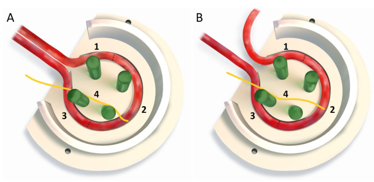
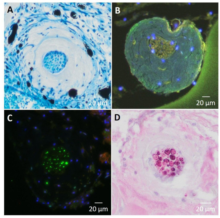
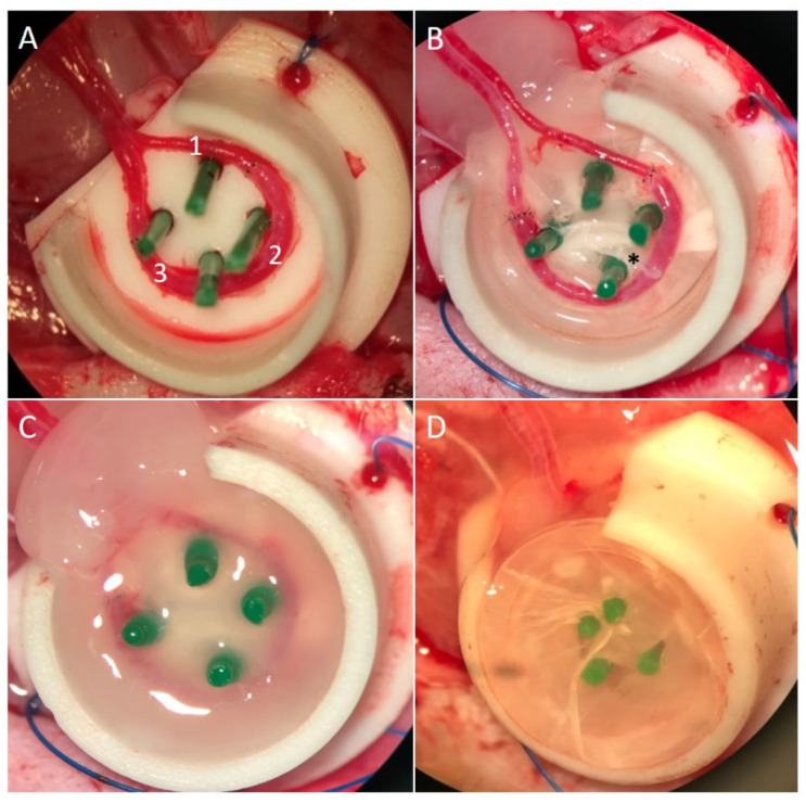
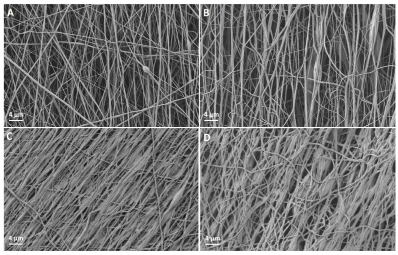
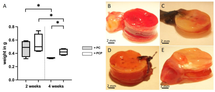
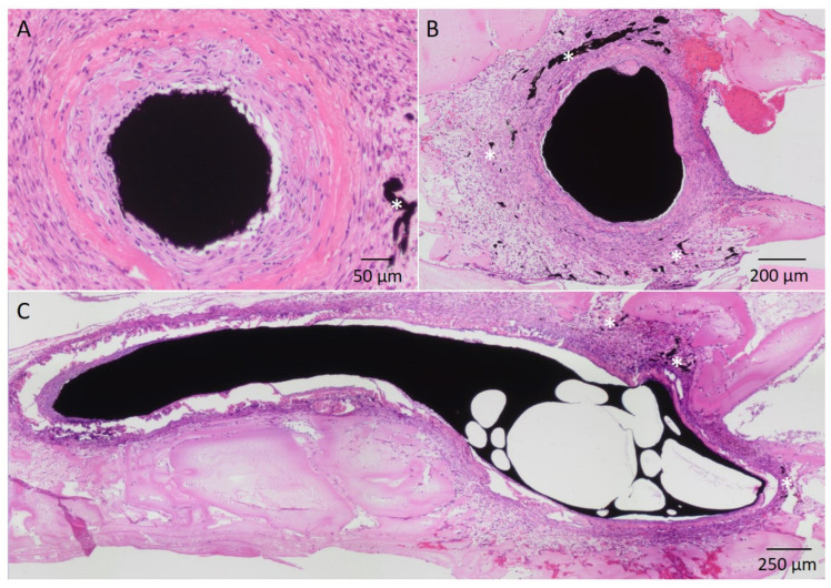
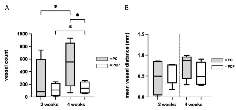
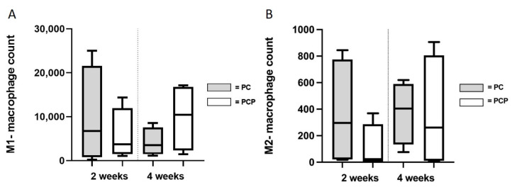
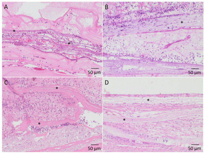
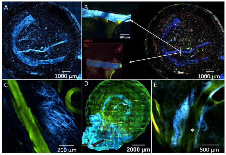
Electrospun nanofibers represent an ideal matrix for the purpose of skeletal muscle tissue engineering due to their highly aligned structure in the nanoscale, mimicking the extracellular matrix of skeletal muscle. However, they often consist of high-density packed fibers, which might impair vascularization. The integration of polyethylene oxide (PEO) sacrificial fibers, which dissolve in water, enables the creation of less dense structures. This study examines potential benefits of poly-ε-caprolactone-collagen I-PEO-nanoscaffolds (PCP) in terms of neovascularization and distribution of newly formed vessels compared to poly-ε-caprolactone -collagen I-nanoscaffolds (PC) in a modified arteriovenous loop model in the rat. For this purpose, the superficial inferior epigastric artery and vein as well as a motor nerve branch were integrated into a multilayer three-dimensional nanofiber scaffold construct, which was enclosed by an isolation chamber. Numbers and spatial distribution of sprouting vessels as well as macrophages were analyzed via immunohistochemistry after two and four weeks of implantation. After four weeks, aligned PC showed a higher number of newly formed vessels, regardless of the compartments formed in PCP by the removal of sacrificial fibers. Both groups showed cell influx and no difference in macrophage invasion. In this study, a model of combined axial vascularization and neurotization of a PCL-collagen I-nanofiber construct could be established for the first time. These results provide a foundation for the in vivo implantation of cells, taking a major step towards the generation of functional skeletal muscle tissue.
电纺纳米纤维因其在纳米尺度上高度排列的结构,类似于骨骼肌的细胞外基质,是骨骼肌组织工程的理想基质。然而,它们通常由高密度的纤维组成,这可能会影响血管生成。聚氧化乙烯(PEO)牺牲纤维的整合,这些纤维可溶于水,可制造出密度较低的结构。本研究通过在大鼠改良动静脉环模型中,比较聚 -ε-己内酯-胶原蛋白 I-PEO-纳米支架(PCP)与聚 -ε-己内酯-胶原蛋白 I-纳米支架(PC)在血管新生和新形成的血管分布方面的潜在益处。为此,将腹壁浅动静脉和一条运动神经分支整合到一个多层三维纳米纤维支架结构中,该结构被一个隔离室包裹。在植入后两周和四周,通过免疫组织化学分析发芽血管和巨噬细胞的数量和空间分布。四周后,无论 PCP 中通过去除牺牲纤维形成的腔室如何,排列整齐的 PC 都显示出更多的新形成的血管。两组均显示细胞浸润,巨噬细胞浸润无差异。在这项研究中,首次成功建立了一种联合轴向血管化和神经化 PCL-胶原蛋白 I-纳米纤维结构的模型。这些结果为细胞的体内植入提供了基础,朝着功能性骨骼肌组织的生成迈出了重要一步。