Department of Plastic and Hand Surgery and Laboratory for Tissue Engineering and Regenerative Medicine, University Hospital of Erlangen, Friedrich-Alexander University of Erlangen-Nürnberg (FAU), Krankenhausstraße 12, 91054, Erlangen, Germany.
Interdisciplinary Clinic for Stem Cell Transplantation, University Cancer Center Hamburg (UCCH), 20246, Hamburg, Germany.
BMC Biotechnol. 2018 Nov 26;18(1):75. doi: 10.1186/s12896-018-0482-6.
The creation of functional skeletal muscle via tissue engineering holds great promise without sacrificing healthy donor tissue. Different cell types have been investigated regarding their myogenic differentiation potential under the influence of various media supplemented with growth factors. Yet, most cell cultures include the use of animal sera, which raises safety concerns and might lead to variances in results. Electrospun nanoscaffolds represent suitable matrices for tissue engineering of skeletal muscle, combining both biocompatibility and stability. We therefore aimed to develop a serum-free myogenic differentiation medium for the co-culture of primary myoblasts (Mb) and mesenchymal stromal cells derived from the bone marrow (BMSC) and adipose tissue (ADSC) on electrospun poly-ε-caprolacton (PCL)-collagen I-nanofibers.
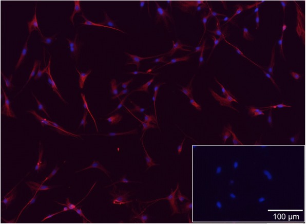
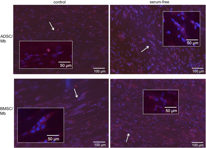
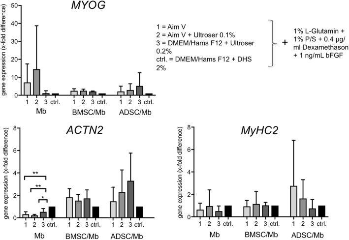
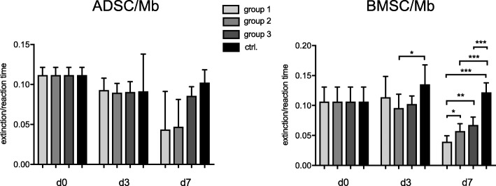
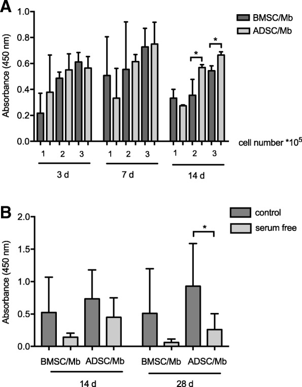
Rat Mb were co-cultured with rat BMSC (BMSC/Mb) or ADSC (ADSC/Mb) two-dimensionally (2D) as monolayers or three-dimensionally (3D) on aligned PCL-collagen I-nanofibers. Differentiation media contained either AIM V, AIM V and Ultroser® G, DMEM/Ham's F12 and Ultroser® G, or donor horse serum (DHS) as a conventional differentiation medium. In 2D co-culture groups, highest upregulation of myogenic markers could be induced by serum-free medium containing DMEM/Ham's F12 and Ultroser® G (group 3) after 7 days. Alpha actinin skeletal muscle 2 (ACTN2) was upregulated 3.3-fold for ADSC/Mb and 1.7-fold for BMSC/Mb after myogenic induction by group 3 serum-free medium when compared to stimulation with DHS. Myogenin (MYOG) was upregulated 5.2-fold in ADSC/Mb and 2.1-fold in BMSC/Mb. On PCL-collagen I-nanoscaffolds, ADSC showed a higher cell viability compared to BMSC in co-culture with Mb. Myosin heavy chain 2, ACTN2, and MYOG as late myogenic markers, showed higher gene expression after long term stimulation with DHS compared to serum-free stimulation, especially in BMSC/Mb co-cultures. Immunocytochemical staining with myosin heavy chain verified the presence of a contractile apparatus under both serum free and standard differentiation conditions.
In this study, we were able to myogenically differentiate mesenchymal stromal cells with myoblasts on PCL-collagen I-nanoscaffolds in a serum-free medium. Our results show that this setting can be used for skeletal muscle tissue engineering, applicable to future clinical applications since no xenogenous substances were used.
通过组织工程创建功能性骨骼肌具有很大的潜力,而无需牺牲健康的供体组织。不同的细胞类型已在不同的培养基中进行了研究,这些培养基中添加了生长因子,以研究其成肌分化潜力。然而,大多数细胞培养都使用动物血清,这引起了安全性问题,并可能导致结果出现差异。静电纺纳米支架是骨骼肌组织工程的合适基质,兼具生物相容性和稳定性。因此,我们旨在开发一种无血清的成肌分化培养基,用于将原代成肌细胞(Mb)与骨髓来源的间充质基质细胞(BMSC)和脂肪组织来源的间充质基质细胞(ADSC)共培养在静电纺聚己内酯(PCL)-I 型胶原纳米纤维上。
将大鼠 Mb 与大鼠 BMSC(BMSC/Mb)或 ADSC(ADSC/Mb)二维(2D)共培养为单层或三维(3D)共培养在排列的 PCL-胶原 I 纳米纤维上。分化培养基中含有 AIM V、AIM V 和 Ultroser® G、DMEM/Ham's F12 和 Ultroser® G 或马血清(DHS)作为传统分化培养基。在 2D 共培养组中,7 天后,含 DMEM/Ham's F12 和 Ultroser® G 的无血清培养基可诱导最高的成肌标志物上调(第 3 组)。与 DHS 刺激相比,第 3 组无血清培养基诱导成肌后,ADSC/Mb 的肌动蛋白 2(ACTN2)上调 3.3 倍,BMSC/Mb 上调 1.7 倍。Myogenin(MYOG)在 ADSC/Mb 中上调 5.2 倍,在 BMSC/Mb 中上调 2.1 倍。在 PCL-胶原 I 纳米支架上,ADSC 在与 Mb 的共培养中比 BMSC 具有更高的细胞活力。肌球蛋白重链 2、ACTN2 和 MYOG 作为晚期成肌标志物,在长期用 DHS 刺激后,其基因表达高于无血清刺激,尤其是在 BMSC/Mb 共培养中。用肌球蛋白重链免疫细胞化学染色证实,在无血清和标准分化条件下均存在收缩装置。
在这项研究中,我们能够在无血清培养基中将间充质基质细胞与成肌细胞共培养在 PCL-胶原 I 纳米支架上进行成肌分化。我们的结果表明,这种设置可用于骨骼肌组织工程,适用于未来的临床应用,因为未使用异种物质。