Human Brain Tissue Bank, Semmelweis University, 1094 Budapest, Hungary.
Human Brain Tissue Bank and Microdissection Laboratory, Semmelweis University, 1094 Budapest, Hungary.
Int J Mol Sci. 2022 Dec 15;23(24):15945. doi: 10.3390/ijms232415945.
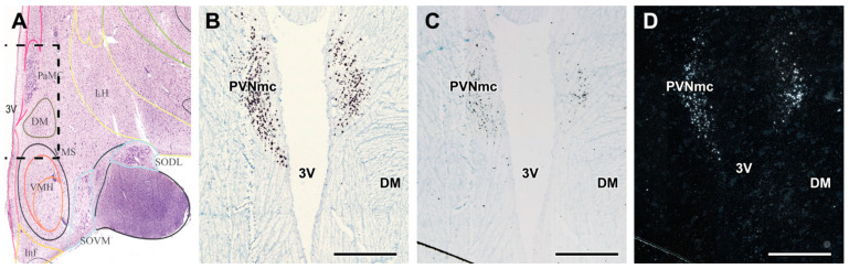
Glucagon-like peptide-1 (GLP-1) receptor (GLP-1R) agonists have been approved for the treatment of type 2 diabetes mellitus (T2DM); however, the brain actions of these drugs are not properly established. We used post mortem microdissected human hypothalamic samples for RT-qPCR and Western blotting. For in situ hybridization histochemistry and immunolabelling, parallel cryosections were prepared from the hypothalamus. We developed in situ hybridization probes for human GLP-1R and oxytocin. In addition, GLP-1 and oxytocin were visualized by immunohistochemistry. Radioactive in situ hybridization histochemistry revealed abundant GLP-1R labelling in the human paraventricular hypothalamic nucleus (PVN), particularly in its magnocellular subdivision (PVNmc). Quantitative analysis of the mRNA signal demonstrated increased GLP-1R expression in the PVNmc in post mortem hypothalamic samples from T2DM subjects as compared to controls, while there was no difference in the expression level of GLP-1R in the other subdivisions of the PVN, the hypothalamic dorsomedial and infundibular nuclei. Our results in the PVN were confirmed by RT-qPCR. Furthermore, we demonstrated by Western blot technique that the GLP-1R protein level was also elevated in the PVN of T2DM patients. GLP-1 fibre terminals were also observed in the PVNmc closely apposing oxytocin neurons using immunohistochemistry. The data suggest that GLP-1 activates GLP-1Rs in the PVNmc and that GLP-1R is elevated in T2DM patients, which may be related to the dysregulation of feeding behaviour and glucose homeostasis in T2DM.
胰高血糖素样肽-1(GLP-1)受体(GLP-1R)激动剂已被批准用于治疗 2 型糖尿病(T2DM);然而,这些药物在大脑中的作用尚未得到充分证实。我们使用死后微解剖的人下丘脑样本进行 RT-qPCR 和 Western blot 分析。为了进行原位杂交组织化学和免疫标记,我们从下丘脑制备了平行的冷冻切片。我们开发了用于人 GLP-1R 和催产素的原位杂交探针。此外,通过免疫组织化学显示 GLP-1 和催产素。放射性原位杂交组织化学显示 GLP-1R 在人室旁下丘脑核(PVN)中大量标记,特别是在其大细胞亚区(PVNmc)中。定量分析 mRNA 信号表明,与对照组相比,T2DM 患者死后下丘脑样本中 PVNmc 中的 GLP-1R 表达增加,而 PVN 的其他亚区、下丘脑背内侧核和漏斗核中 GLP-1R 的表达水平没有差异。我们在 PVN 中的结果通过 RT-qPCR 得到了证实。此外,我们还通过 Western blot 技术证明,T2DM 患者的 PVN 中 GLP-1R 蛋白水平也升高了。使用免疫组织化学还观察到 GLP-1 纤维末端紧密毗邻催产素神经元存在于 PVNmc 中。这些数据表明,GLP-1 在 PVNmc 中激活 GLP-1R,并且在 T2DM 患者中 GLP-1R 升高,这可能与 T2DM 中摄食行为和葡萄糖稳态的失调有关。