Department of Cardiothoracic Surgery, University Hospitals Coventry and Warwickshire NHS Trust, Coventry CV2 2DX, UK.
Laboratory of Experimental Surgery and Surgical Research "N. S. Christeas", Medical School, National and Kapodistrian University of Athens, 11527 Athens, Greece.
Medicina (Kaunas). 2022 Dec 15;58(12):1842. doi: 10.3390/medicina58121842.
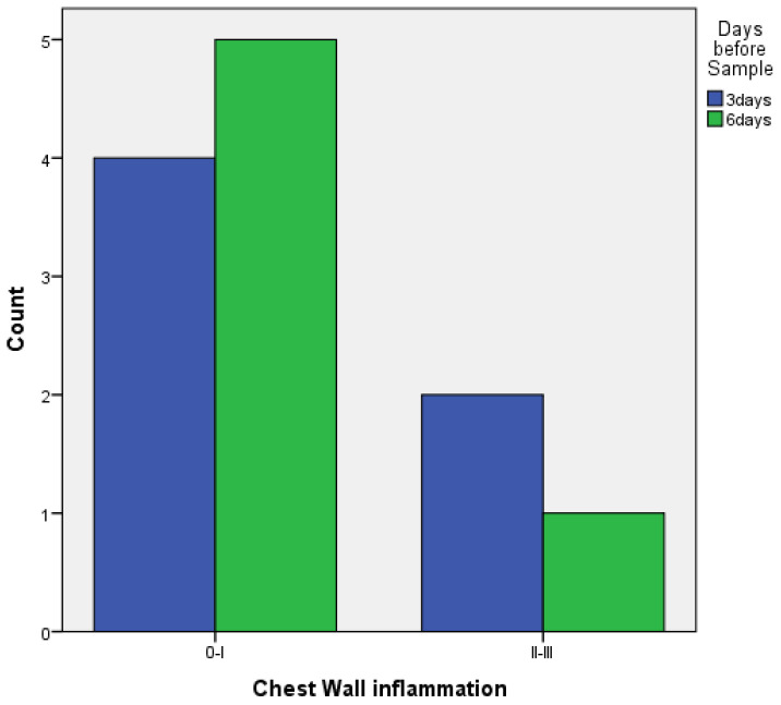
: This study was designed to evaluate platelet-rich plasma (PRP) as a method of pleurodesis in a rabbit model. Pleurodesis with PRP was compared against the gold-standard use of talc. The secondary evaluation assessed the ideal time for achieving pleurodesis. : 25 healthy New Zealand white rabbits were assigned to three groups, as follows: 12 animals in the first and second groups, as well as one animal with no intervention in the final group, which was used as a control. The talc pleurodesis group (baseline) underwent pleurodesis with sterile talc, which is the gold-standard sclerosing agent used for pleurodesis. The PRP group underwent pleurodesis using autologous PRP. The last group had one rabbit with no intervention. A total of 12 rabbits ( = 6 for the talc pleurodesis group and = 6 for the PRP group) were sacrificed 3 days (72 h) after the intervention, and 12 rabbits ( = 6 for the talc pleurodesis group and = 6 for the PRP group) were sacrificed 6 days (144 h) after the intervention. In both the talc and PRP group, FBC and CRP were measured before the intervention and in 3 or 6 days afterwards, respectively. The pleura and the lungs were evaluated histopathologically. : Macroscopically, there were no statistically significant differences between the two groups. In terms of microscopic findings, there were no statistically significant differences in inflammatory reactions provoked in the visceral and parietal pleura between the PRP and talc. In addition, with talc pleurodesis, a foreign-body reaction was observed in about 50% of the cases, which was not observed with PRP. In terms of inflammation between 3 and 6 days, there were no statistically significant differences with PRP, there was only a statistically significant difference between 3 and 6 days regarding the parietal pleura in the talc group. : The instillation of autologous PRP in the pleural cavity shows promise in achieving pleurodesis. The efficacy of PRP as a pleurodesis agent should be examined further.
: 本研究旨在评估富血小板血浆(PRP)作为一种兔胸膜固定术方法。将 PRP 胸膜固定术与滑石粉的金标准使用进行比较。次要评估评估了实现胸膜固定术的理想时间。 : 25 只健康的新西兰白兔被分为三组,如下所示:第一组和第二组各 12 只动物,最后一组 1 只动物无干预,作为对照。滑石粉胸膜固定术组(基线)接受无菌滑石粉胸膜固定术,滑石粉是用于胸膜固定术的金标准硬化剂。PRP 组接受自体 PRP 胸膜固定术。最后一组有一只兔子没有干预。共有 12 只兔子(滑石粉胸膜固定术组 6 只,PRP 组 6 只)在干预后 3 天(72 小时)被处死,另有 12 只兔子(滑石粉胸膜固定术组 6 只,PRP 组 6 只)在干预后 6 天(144 小时)被处死。在滑石粉组和 PRP 组中,FBC 和 CRP 分别在干预前和干预后 3 天或 6 天测量。胸膜和肺进行组织病理学评估。 : 大体上,两组之间没有统计学上的显著差异。在微观发现方面,PRP 和滑石粉在脏层和壁层胸膜引起的炎症反应方面没有统计学上的显著差异。此外,在滑石粉胸膜固定术中,约 50%的病例观察到异物反应,而 PRP 则没有观察到。在 3 天至 6 天的炎症方面,PRP 之间没有统计学上的显著差异,仅在滑石粉组的壁层胸膜方面,3 天和 6 天之间存在统计学上的显著差异。 : 自体 PRP 胸腔内灌注在实现胸膜固定术中显示出前景。PRP 作为胸膜固定剂的疗效应进一步研究。