• 文献检索
  • 文档翻译
  • 深度研究
  • 学术资讯
  • Suppr Zotero 插件Zotero 插件
  • 邀请有礼
  • 套餐&价格
  • 历史记录
应用&插件
Suppr Zotero 插件Zotero 插件浏览器插件Mac 客户端Windows 客户端微信小程序
定价
高级版会员购买积分包购买API积分包
服务
文献检索文档翻译深度研究API 文档MCP 服务
关于我们
关于 Suppr公司介绍联系我们用户协议隐私条款
关注我们

Suppr 超能文献

核心技术专利:CN118964589B侵权必究
粤ICP备2023148730 号-1Suppr @ 2026

文献检索

告别复杂PubMed语法,用中文像聊天一样搜索,搜遍4000万医学文献。AI智能推荐,让科研检索更轻松。

立即免费搜索

文件翻译

保留排版,准确专业,支持PDF/Word/PPT等文件格式,支持 12+语言互译。

免费翻译文档

深度研究

AI帮你快速写综述,25分钟生成高质量综述,智能提取关键信息,辅助科研写作。

立即免费体验

基于 ImageJ 的工具,用于不同类型的显微镜图像之间的三维配准。

An ImageJ-based tool for three-dimensional registration between different types of microscopic images.

机构信息

Division of Embryology, National Institute for Basic Biology, Okazaki, Japan.

Department of Basic Biology, School of Life Science, SOKENDAI (The Graduate University for Advanced Studies), Okazaki, Japan.

出版信息

Dev Growth Differ. 2023 Jan;65(1):65-74. doi: 10.1111/dgd.12835. Epub 2023 Jan 14.

DOI:10.1111/dgd.12835
PMID:36576380
原文链接:https://pmc.ncbi.nlm.nih.gov/articles/PMC10107647/
Abstract

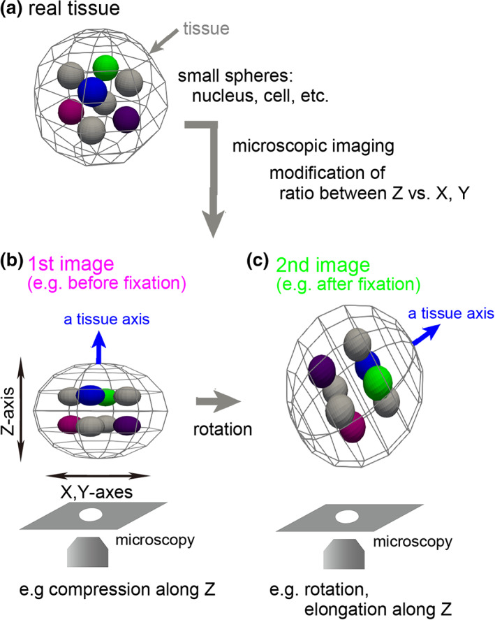
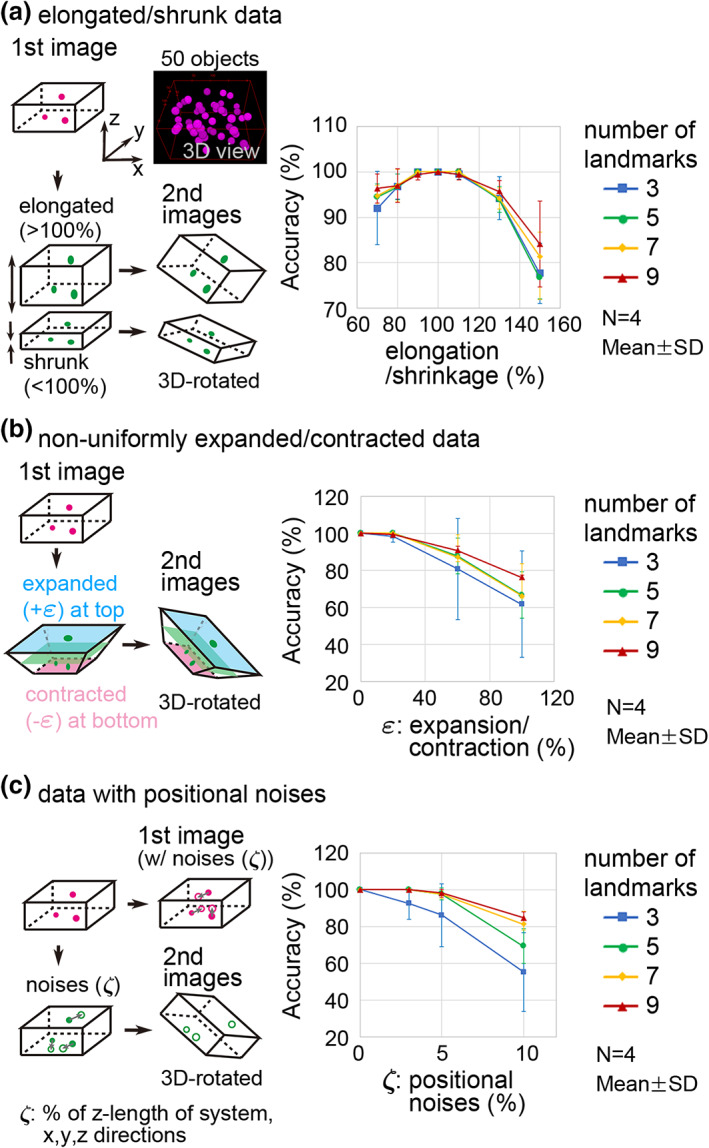
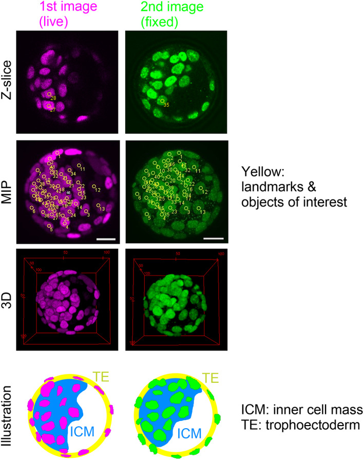
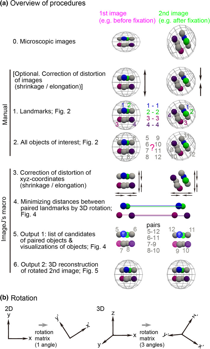
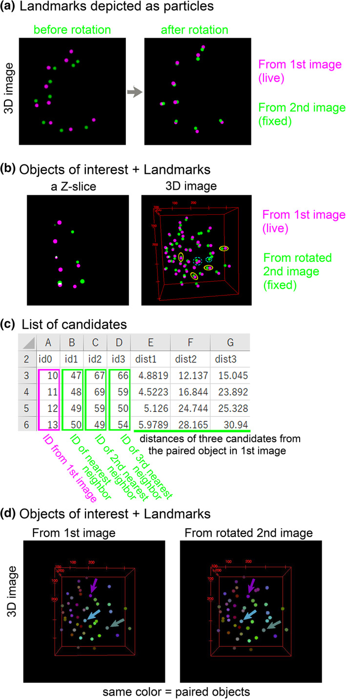
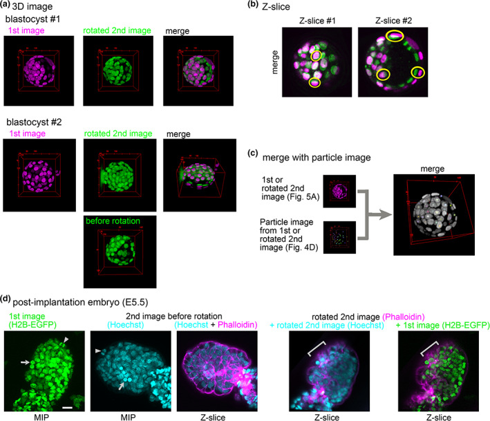
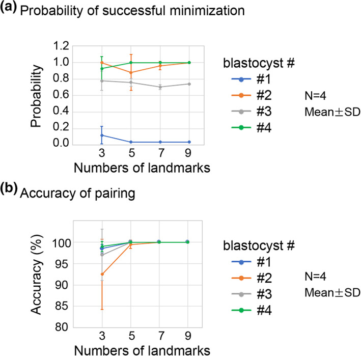
Three-dimensional (3D) registration (i.e., alignment) between two microscopic images is very helpful to study tissues that do not adhere to substrates, such as mouse embryos and organoids, which are often 3D rotated during imaging. However, there is no 3D registration tool easily accessible for experimental biologists. Here we developed an ImageJ-based tool which allows for 3D registration accompanied with both quantitative evaluation of the accuracy and reconstruction of 3D rotated images. In this tool, several landmarks are manually provided in two images to be aligned, and 3D rotation is computed so that the distances between the paired landmarks from the two images are minimized. By simultaneously providing multiple points (e.g., all nuclei in the regions of interest) other than the landmarks in the two images, the correspondence of each point between the two images, i.e., to which nucleus in one image a certain nucleus in another image corresponds, is quantitatively explored. Furthermore, 3D rotation is applied to one of the two images, resulting in reconstruction of 3D rotated images. We demonstrated that this tool successfully achieved 3D registration and reconstruction of images in mouse pre- and post-implantation embryos, where one image was obtained during live imaging and another image was obtained from fixed embryos after live imaging. This approach provides a versatile tool applicable for various tissues and species.

摘要

三维(3D)配准(即对齐)在研究不附着于基质的组织(如经常在成像过程中 3D 旋转的小鼠胚胎和类器官)时非常有帮助。然而,对于实验生物学家来说,没有易于使用的 3D 配准工具。在这里,我们开发了一种基于 ImageJ 的工具,它允许进行 3D 配准,并伴随对准确性的定量评估和 3D 旋转图像的重建。在这个工具中,在要对齐的两个图像中手动提供几个标记点,并计算 3D 旋转,使得来自两个图像的成对标记点之间的距离最小化。通过同时在两个图像中提供多个点(例如,感兴趣区域中的所有核)而不是标记点,可以定量探索两个图像之间每个点的对应关系,即一个图像中的某个核与另一个图像中的哪个核相对应。此外,将 3D 旋转应用于两个图像中的一个图像,从而重建 3D 旋转图像。我们证明了该工具成功地实现了小鼠植入前和植入后胚胎的 3D 配准和重建,其中一个图像是在活体成像过程中获得的,另一个图像是在活体成像后从固定胚胎中获得的。这种方法提供了一种适用于各种组织和物种的通用工具。

https://cdn.ncbi.nlm.nih.gov/pmc/blobs/efa1/10107647/b4cf48ab17a0/DGD-65-65-g008.jpg
https://cdn.ncbi.nlm.nih.gov/pmc/blobs/efa1/10107647/589017e297b2/DGD-65-65-g006.jpg
https://cdn.ncbi.nlm.nih.gov/pmc/blobs/efa1/10107647/7c75d062c8e4/DGD-65-65-g002.jpg
https://cdn.ncbi.nlm.nih.gov/pmc/blobs/efa1/10107647/05ed24954fc8/DGD-65-65-g005.jpg
https://cdn.ncbi.nlm.nih.gov/pmc/blobs/efa1/10107647/b74ff5711629/DGD-65-65-g003.jpg
https://cdn.ncbi.nlm.nih.gov/pmc/blobs/efa1/10107647/baa9e05a36a0/DGD-65-65-g001.jpg
https://cdn.ncbi.nlm.nih.gov/pmc/blobs/efa1/10107647/50b4336e3ab0/DGD-65-65-g007.jpg
https://cdn.ncbi.nlm.nih.gov/pmc/blobs/efa1/10107647/b4cf48ab17a0/DGD-65-65-g008.jpg

相似文献

1
An ImageJ-based tool for three-dimensional registration between different types of microscopic images.基于 ImageJ 的工具,用于不同类型的显微镜图像之间的三维配准。
Dev Growth Differ. 2023 Jan;65(1):65-74. doi: 10.1111/dgd.12835. Epub 2023 Jan 14.
2
Automatic three-dimensional segmentation of mouse embryonic stem cell nuclei by utilising multiple channels of confocal fluorescence images.利用共聚焦荧光图像的多个通道自动对小鼠胚胎干细胞核进行三维分割。
J Microsc. 2021 Jan;281(1):57-75. doi: 10.1111/jmi.12949. Epub 2020 Aug 8.
3
Registration of serial sections of mouse liver cell nuclei.小鼠肝细胞核连续切片的登记
J Microsc. 1998 Oct;192(Pt 1):37-53. doi: 10.1046/j.1365-2818.1998.00405.x.
4
Three-dimensional quantitative evaluation method of nonrigid registration algorithms for adaptive radiotherapy.自适应放疗中用于非刚性配准算法的三维定量评估方法。
Med Phys. 2010 Mar;37(3):1137-45. doi: 10.1118/1.3302916.
5
Evaluating the utility of intraprocedural 3D TRUS image information in guiding registration for displacement compensation during prostate biopsy.评估术中三维超声(3D TRUS)图像信息在前列腺活检期间引导位移补偿配准中的效用。
Med Phys. 2014 Aug;41(8):082901. doi: 10.1118/1.4885959.
6
Position tracking of moving liver lesion based on real-time registration between 2D ultrasound and 3D preoperative images.基于二维超声与三维术前图像实时配准的肝脏移动病灶位置跟踪
Med Phys. 2015 Jan;42(1):335-47. doi: 10.1118/1.4903945.
7
Improving superficial target delineation in radiation therapy with endoscopic tracking and registration.利用内镜跟踪和配准技术改善放射治疗中的表面靶区勾画。
Med Phys. 2011 Dec;38(12):6458-68. doi: 10.1118/1.3658569.
8
Thorax x-ray and CT interventional dataset for nonrigid 2D/3D image registration evaluation.用于非刚性 2D/3D 图像配准评估的胸部 X 射线和 CT 介入数据集。
Med Phys. 2018 Nov;45(11):5343-5351. doi: 10.1002/mp.13174. Epub 2018 Oct 1.
9
Assessment of image registration accuracy in three-dimensional transrectal ultrasound guided prostate biopsy.三维经直肠超声引导前列腺穿刺活检中图像配准精度的评估。
Med Phys. 2010 Feb;37(2):802-13. doi: 10.1118/1.3298010.
10
A generic classification-based method for segmentation of nuclei in 3D images of early embryos.基于分类的通用方法,用于分割早期胚胎 3D 图像中的细胞核。
BMC Bioinformatics. 2014 Jan 14;15:9. doi: 10.1186/1471-2105-15-9.

本文引用的文献

1
Effective mechanical potential of cell-cell interaction explains three-dimensional morphologies during early embryogenesis.细胞间相互作用的有效力学势解释了早期胚胎发生过程中的三维形态。
PLoS Comput Biol. 2023 Aug 7;19(8):e1011306. doi: 10.1371/journal.pcbi.1011306. eCollection 2023 Aug.
2
QuantifyPolarity, a new tool-kit for measuring planar polarized protein distributions and cell properties in developing tissues.量化偏振度,一种用于测量发育组织中平面偏振蛋白分布和细胞特性的新工具包。
Development. 2021 Sep 1;148(18). doi: 10.1242/dev.198952. Epub 2021 Sep 7.
3
3D time-lapse microscopy paired with endpoint lineage analysis in mouse blastocysts.
3D 延时显微镜与小鼠囊胚终点谱系分析相结合。
STAR Protoc. 2021 Apr 8;2(2):100446. doi: 10.1016/j.xpro.2021.100446. eCollection 2021 Jun 18.
4
Live Visualization of ERK Activity in the Mouse Blastocyst Reveals Lineage-Specific Signaling Dynamics.在小鼠囊胚中实时可视化 ERK 活性揭示谱系特异性信号动力学。
Dev Cell. 2020 Nov 9;55(3):341-353.e5. doi: 10.1016/j.devcel.2020.09.030. Epub 2020 Oct 21.
5
Cell-Cycle-Dependent ERK Signaling Dynamics Direct Fate Specification in the Mammalian Preimplantation Embryo.细胞周期依赖性 ERK 信号动态直接决定哺乳动物着床前胚胎的命运。
Dev Cell. 2020 Nov 9;55(3):328-340.e5. doi: 10.1016/j.devcel.2020.09.013. Epub 2020 Oct 21.
6
Fijiyama: a registration tool for 3D multimodal time-lapse imaging.菲几雅玛:三维多时相延时成像的注册工具。
Bioinformatics. 2021 Jun 16;37(10):1482-1484. doi: 10.1093/bioinformatics/btaa846.
7
A chordate species lacking utilizes calcium oscillation and for left-right patterning.一种缺乏 的脊索动物物种利用钙震荡和 进行左右模式形成。
Proc Natl Acad Sci U S A. 2020 Feb 25;117(8):4188-4198. doi: 10.1073/pnas.1916858117. Epub 2020 Feb 6.
8
In Toto Imaging and Reconstruction of Post-Implantation Mouse Development at the Single-Cell Level.单细胞水平下的植入后小鼠发育的全胚胎成像和重建。
Cell. 2018 Oct 18;175(3):859-876.e33. doi: 10.1016/j.cell.2018.09.031. Epub 2018 Oct 11.
9
Biologically constrained optimization based cell membrane segmentation in C. elegans embryos.基于生物约束优化的秀丽隐杆线虫胚胎细胞膜分割
BMC Bioinformatics. 2017 Jun 19;18(1):307. doi: 10.1186/s12859-017-1717-6.
10
EpiTools: An Open-Source Image Analysis Toolkit for Quantifying Epithelial Growth Dynamics.EpiTools:用于量化上皮生长动力学的开源图像分析工具包。
Dev Cell. 2016 Jan 11;36(1):103-116. doi: 10.1016/j.devcel.2015.12.012.