Lin Li, Tong Xin, Cavallero Susana, Zhang Yide, Na Shuai, Cao Rui, Hsiai Tzung K, Wang Lihong V
Caltech Optical Imaging Laboratory, Andrew and Peggy Cherng Department of Medical Engineering, Department of Electrical Engineering, California Institute of Technology, Pasadena, CA, USA.
College of Biomedical Engineering and Instrument Science, Zhejiang University, Hangzhou, China.
Light Sci Appl. 2023 Jan 3;12(1):12. doi: 10.1038/s41377-022-01053-7.
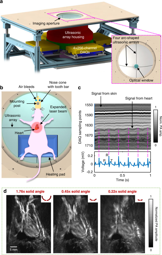
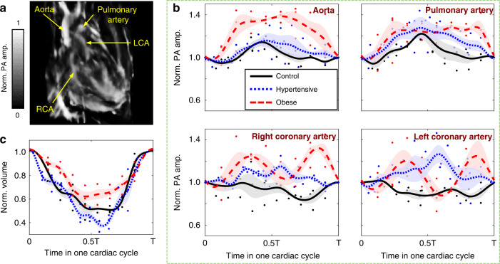
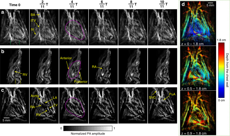
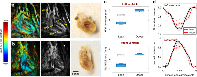
Complementary to mainstream cardiac imaging modalities for preclinical research, photoacoustic computed tomography (PACT) can provide functional optical contrast with high imaging speed and resolution. However, PACT has not been demonstrated to reveal the dynamics of whole cardiac anatomy or vascular system without surgical procedure (thoracotomy) for tissue penetration. Here, we achieved non-invasive imaging of rat hearts using the recently developed three-dimensional PACT (3D-PACT) platform, demonstrating the regulated illumination and detection schemes to reduce the effects of optical attenuation and acoustic distortion through the chest wall; thereby, enabling unimpeded visualization of the cardiac anatomy and intracardiac hemodynamics following rapidly scanning the heart within 10 s. We further applied 3D-PACT to reveal distinct cardiac structural and functional changes among the healthy, hypertensive, and obese rats, with optical contrast to uncover differences in cardiac chamber size, wall thickness, and hemodynamics. Accordingly, 3D-PACT provides high imaging speed and nonionizing penetration to capture the whole heart for diagnosing the animal models, holding promises for clinical translation to cardiac imaging of human neonates.
作为临床前研究中主流心脏成像模式的补充,光声计算机断层扫描(PACT)能够以高成像速度和分辨率提供功能性光学对比度。然而,在不进行用于组织穿透的外科手术(开胸术)的情况下,PACT尚未被证明能够揭示整个心脏解剖结构或血管系统的动态变化。在此,我们使用最近开发的三维PACT(3D-PACT)平台实现了对大鼠心脏的无创成像,展示了通过调节照明和检测方案来减少光衰减和声失真对穿过胸壁的影响;从而,能够在10秒内快速扫描心脏后,清晰地观察到心脏解剖结构和心内血流动力学。我们进一步应用3D-PACT揭示健康、高血压和肥胖大鼠之间不同的心脏结构和功能变化,并利用光学对比度发现心腔大小、壁厚和血流动力学方面的差异。因此,3D-PACT提供了高成像速度和非电离穿透能力,能够对整个心脏进行成像以诊断动物模型,有望转化应用于人类新生儿心脏成像的临床研究。