State Key Laboratory of Surface Physics and Department of Physics, Human Phenome Institute, Academy for Engineering and Technology, Key Laboratory of Micro and Nano Photonic Structures (Ministry of Education), Yiwu Research Institute of Fudan University, Fudan University, Shanghai, P.R. China.
Department of Urology, Ren Ji Hospital, School of Medicine, Shanghai Jiao Tong University, Shanghai, P.R. China.
Cancer Res. 2023 Feb 15;83(4):641-651. doi: 10.1158/0008-5472.CAN-22-2146.
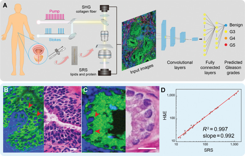
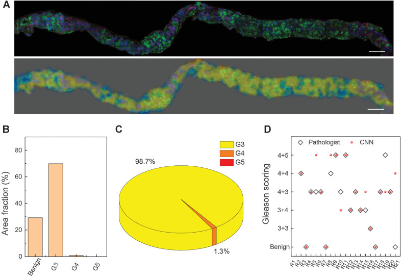
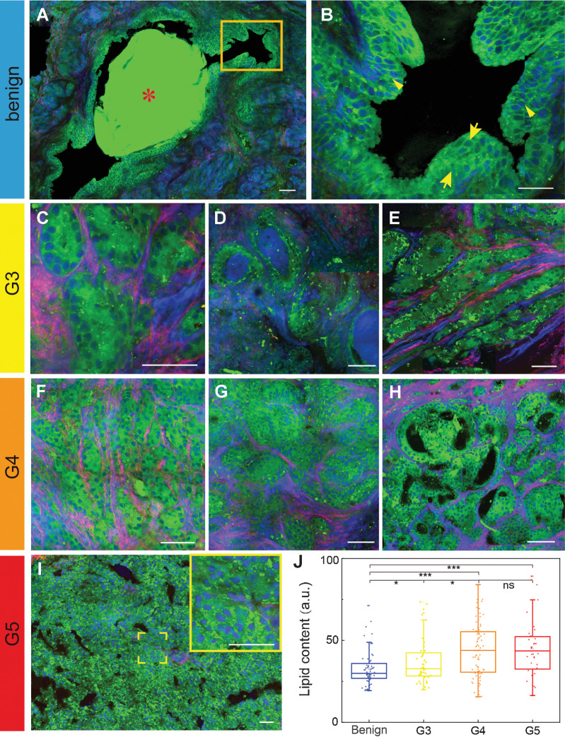
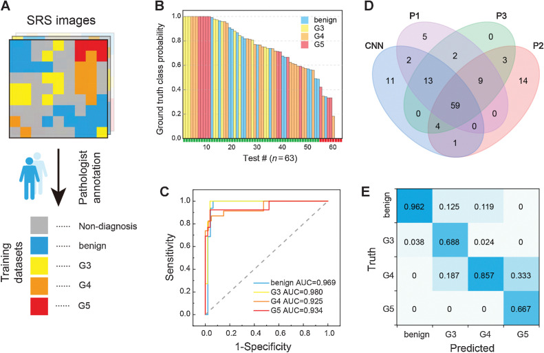
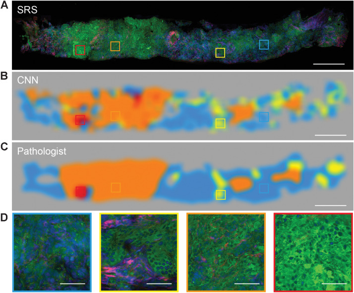
Focal therapy (FT) has been proposed as an approach to eradicate clinically significant prostate cancer while preserving the normal surrounding tissues to minimize treatment-related toxicity. Rapid histology of core needle biopsies is essential to ensure the precise FT for localized lesions and to determine tumor grades. However, it is difficult to achieve both high accuracy and speed with currently available histopathology methods. Here, we demonstrated that stimulated Raman scattering (SRS) microscopy could reveal the largely heterogeneous histologic features of fresh prostatic biopsy tissues in a label-free and near real-time manner. A diagnostic convolutional neural network (CNN) built based on images from 61 patients could classify Gleason patterns of prostate cancer with an accuracy of 85.7%. An additional 22 independent cases introduced as external test dataset validated the CNN performance with 84.4% accuracy. Gleason scores of core needle biopsies from 21 cases were calculated using the deep learning SRS system and showed a 71% diagnostic consistency with grading from three pathologists. This study demonstrates the potential of a deep learning-assisted SRS platform in evaluating the tumor grade of prostate cancer, which could help simplify the diagnostic workflow and provide timely histopathology compatible with FT treatment.
A platform combining stimulated Raman scattering microscopy and a convolutional neural network provides rapid histopathology and automated Gleason scoring on fresh prostate core needle biopsies without complex tissue processing.
焦点治疗(FT)已被提议作为一种方法,以根除临床上显著的前列腺癌,同时保留正常的周围组织,以最大程度地减少与治疗相关的毒性。核心针活检的快速组织学对于确保对局部病变进行精确的 FT 以及确定肿瘤分级至关重要。然而,目前可用的组织病理学方法很难同时实现高准确性和速度。在这里,我们证明了受激拉曼散射(SRS)显微镜可以无标记且近乎实时地揭示新鲜前列腺活检组织的大异质组织学特征。基于 61 名患者的图像构建的诊断卷积神经网络(CNN)可以以 85.7%的准确率对前列腺癌的 Gleason 模式进行分类。另外 22 个独立的案例作为外部测试数据集引入,以 84.4%的准确率验证了 CNN 的性能。使用深度学习 SRS 系统计算了 21 例核心针活检的 Gleason 评分,与三位病理学家的分级相比具有 71%的诊断一致性。本研究证明了深度学习辅助 SRS 平台在评估前列腺癌肿瘤分级方面的潜力,这有助于简化诊断工作流程并提供与 FT 治疗兼容的及时组织病理学。
结合受激拉曼散射显微镜和卷积神经网络的平台可对新鲜前列腺核心针活检进行快速组织病理学和自动 Gleason 评分,而无需复杂的组织处理。