• 文献检索
  • 文档翻译
  • 深度研究
  • 学术资讯
  • Suppr Zotero 插件Zotero 插件
  • 邀请有礼
  • 套餐&价格
  • 历史记录
应用&插件
Suppr Zotero 插件Zotero 插件浏览器插件Mac 客户端Windows 客户端微信小程序
定价
高级版会员购买积分包购买API积分包
服务
文献检索文档翻译深度研究API 文档MCP 服务
关于我们
关于 Suppr公司介绍联系我们用户协议隐私条款
关注我们

Suppr 超能文献

核心技术专利:CN118964589B侵权必究
粤ICP备2023148730 号-1Suppr @ 2026

文献检索

告别复杂PubMed语法,用中文像聊天一样搜索,搜遍4000万医学文献。AI智能推荐,让科研检索更轻松。

立即免费搜索

文件翻译

保留排版,准确专业,支持PDF/Word/PPT等文件格式,支持 12+语言互译。

免费翻译文档

深度研究

AI帮你快速写综述,25分钟生成高质量综述,智能提取关键信息,辅助科研写作。

立即免费体验

应用受激拉曼散射显微镜和多实例学习对未经处理的乳腺芯针活检组织进行组织学诊断。

Histological diagnosis of unprocessed breast core-needle biopsy via stimulated Raman scattering microscopy and multi-instance learning.

机构信息

State Key Laboratory of Surface Physics and Department of Physics, Human Phenome Institute, Academy for Engineering and Technology, Key Laboratory of Micro and Nano Photonic Structures (Ministry of Education), Yiwu Research Institute, Fudan University, Shanghai 200433, China.

Department of Pathology, Fudan University Shanghai Cancer Center, Shanghai 200032, China.

出版信息

Theranostics. 2023 Feb 21;13(4):1342-1354. doi: 10.7150/thno.81784. eCollection 2023.

DOI:10.7150/thno.81784
PMID:36923541
原文链接:https://pmc.ncbi.nlm.nih.gov/articles/PMC10008736/
Abstract

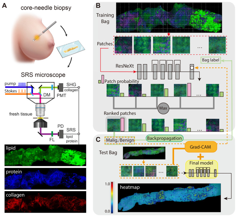
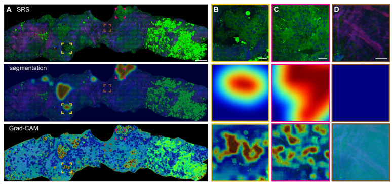
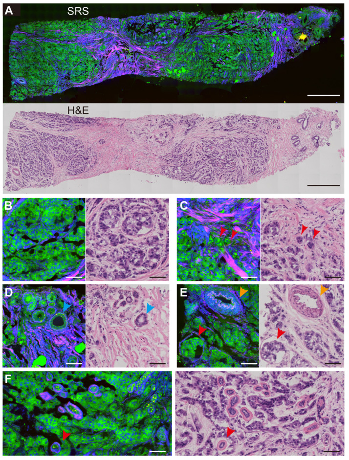
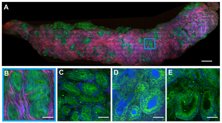
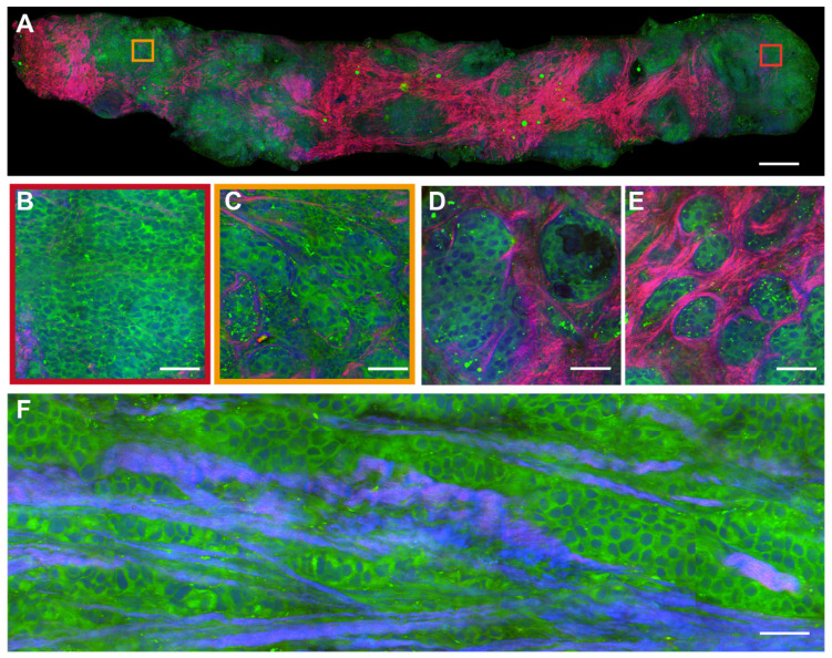
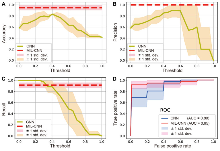
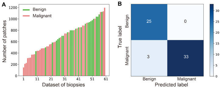
Core-needle biopsy (CNB) plays a vital role in the initial diagnosis of breast cancer. However, the complex tissue processing and global shortage of pathologists have hindered traditional histopathology from timely diagnosis on fresh biopsies. In this work, we developed a full digital platform by integrating label-free stimulated Raman scattering (SRS) microscopy with weakly-supervised learning for rapid and automated cancer diagnosis on un-labelled breast CNB. We first compared the results of SRS imaging with standard hematoxylin and eosin (H&E) staining on adjacent frozen tissue sections. Then fresh unprocessed biopsy tissues were imaged by SRS to reveal diagnostic histoarchitectures. Next, weakly-supervised learning, i.e., the multi-instance learning (MIL) model was conducted to evaluate the ability to differentiate between benign and malignant cases, and compared with the performance of supervised learning model. Finally, gradient-weighted class activation mapping (Grad-CAM) and semantic segmentation were performed to spatially resolve benign/malignant areas with high efficiency. We verified the ability of SRS in revealing essential histological hallmarks of breast cancer in both thin frozen sections and fresh unprocessed biopsy, generating histoarchitectures well correlated with H&E staining. Moreover, we demonstrated that weakly-supervised MIL model could achieve superior classification performance to supervised learnings, reaching diagnostic accuracy of 95% on 61 biopsy specimens. Furthermore, Grad-CAM allowed the trained MIL model to visualize the histological heterogeneity within the CNB. Our results indicate that MIL-assisted SRS microscopy provides rapid and accurate diagnosis on histologically heterogeneous breast CNB, and could potentially help the subsequent management of patients.

摘要

核心针活检 (CNB) 在乳腺癌的初步诊断中起着至关重要的作用。然而,复杂的组织处理和全球病理学家短缺阻碍了传统的组织病理学对新鲜活检进行及时诊断。在这项工作中,我们通过整合无标记的受激拉曼散射 (SRS) 显微镜与弱监督学习,为未标记的乳腺癌 CNB 上的快速和自动癌症诊断开发了一个全数字平台。我们首先比较了 SRS 成像与相邻冷冻组织切片上标准苏木精和伊红 (H&E) 染色的结果。然后,对新鲜未处理的活检组织进行 SRS 成像以揭示诊断性组织架构。接下来,进行弱监督学习,即多实例学习 (MIL) 模型,以评估区分良性和恶性病例的能力,并与监督学习模型的性能进行比较。最后,进行梯度加权类激活映射 (Grad-CAM) 和语义分割,以高效地空间解析良性/恶性区域。我们验证了 SRS 在揭示乳腺癌在薄冷冻切片和新鲜未处理活检中的基本组织学特征的能力,生成的组织架构与 H&E 染色高度相关。此外,我们证明了弱监督 MIL 模型可以达到优于监督学习的分类性能,在 61 个活检标本上达到 95%的诊断准确性。此外,Grad-CAM 允许训练有素的 MIL 模型对 CNB 内的组织学异质性进行可视化。我们的结果表明,MIL 辅助的 SRS 显微镜为组织学异质的乳腺癌 CNB 提供了快速准确的诊断,并且有可能有助于患者的后续管理。

https://cdn.ncbi.nlm.nih.gov/pmc/blobs/842e/10008736/1010e837570f/thnov13p1342g007.jpg
https://cdn.ncbi.nlm.nih.gov/pmc/blobs/842e/10008736/2d5ed9530d0f/thnov13p1342g001.jpg
https://cdn.ncbi.nlm.nih.gov/pmc/blobs/842e/10008736/354d0df72f3b/thnov13p1342g002.jpg
https://cdn.ncbi.nlm.nih.gov/pmc/blobs/842e/10008736/7683151d9245/thnov13p1342g003.jpg
https://cdn.ncbi.nlm.nih.gov/pmc/blobs/842e/10008736/6a6e24822aeb/thnov13p1342g004.jpg
https://cdn.ncbi.nlm.nih.gov/pmc/blobs/842e/10008736/389b9248259a/thnov13p1342g005.jpg
https://cdn.ncbi.nlm.nih.gov/pmc/blobs/842e/10008736/4abf7d7186ba/thnov13p1342g006.jpg
https://cdn.ncbi.nlm.nih.gov/pmc/blobs/842e/10008736/1010e837570f/thnov13p1342g007.jpg

相似文献

1
Histological diagnosis of unprocessed breast core-needle biopsy via stimulated Raman scattering microscopy and multi-instance learning.应用受激拉曼散射显微镜和多实例学习对未经处理的乳腺芯针活检组织进行组织学诊断。
Theranostics. 2023 Feb 21;13(4):1342-1354. doi: 10.7150/thno.81784. eCollection 2023.
2
Rapid histology of laryngeal squamous cell carcinoma with deep-learning based stimulated Raman scattering microscopy.基于深度学习的受激拉曼散射显微镜快速检测喉鳞状细胞癌。
Theranostics. 2019 Apr 13;9(9):2541-2554. doi: 10.7150/thno.32655. eCollection 2019.
3
Stimulated Raman Scattering Microscopy Enables Gleason Scoring of Prostate Core Needle Biopsy by a Convolutional Neural Network.受激拉曼散射显微镜通过卷积神经网络实现前列腺核心针活检的 Gleason 评分。
Cancer Res. 2023 Feb 15;83(4):641-651. doi: 10.1158/0008-5472.CAN-22-2146.
4
Comparative analysis of confocal microscopy on fresh breast core needle biopsies and conventional histology.对比新鲜乳腺芯针活检的共聚焦显微镜检查和常规组织学检查。
Diagn Pathol. 2019 Jun 15;14(1):58. doi: 10.1186/s13000-019-0835-z.
5
Label-Free Histology and Evaluation of Human Pancreatic Cancer with Coherent Nonlinear Optical Microscopy.无标记组织学与相干非线性光学显微镜在人胰腺癌评估中的应用。
Anal Chem. 2021 Nov 23;93(46):15550-15558. doi: 10.1021/acs.analchem.1c03861. Epub 2021 Nov 9.
6
Stimulated Raman histology as a method to determine the adequacy of renal mass biopsy and identify malignant subtypes of renal cell carcinoma.应用受激拉曼组织学确定肾肿瘤穿刺活检标本的充分性并识别肾细胞癌的恶性亚型。
Urol Oncol. 2023 Jul;41(7):328.e9-328.e13. doi: 10.1016/j.urolonc.2023.04.008. Epub 2023 May 23.
7
The accuracy of frozen section analysis in ultrasound- guided core needle biopsy of breast lesions.超声引导下乳腺病变粗针穿刺活检中冰冻切片分析的准确性。
BMC Cancer. 2009 Sep 24;9:341. doi: 10.1186/1471-2407-9-341.
8
DRAQ5 and Eosin ('D&E') as an Analog to Hematoxylin and Eosin for Rapid Fluorescence Histology of Fresh Tissues.DRAQ5与伊红(“D&E”)作为苏木精和伊红的类似物用于新鲜组织的快速荧光组织学检查
PLoS One. 2016 Oct 27;11(10):e0165530. doi: 10.1371/journal.pone.0165530. eCollection 2016.
9
Discrepancies between radiological and histological findings in preoperative core needle (CNB) and vacuum-assisted (VAB) breast biopsies.术前空心针(CNB)和真空辅助(VAB)乳腺活检的影像学和组织学检查结果差异。
J Cancer Res Clin Oncol. 2021 Mar;147(3):749-754. doi: 10.1007/s00432-020-03481-7. Epub 2020 Dec 7.
10
Ultrasound-guided core needle biopsy of the breast: does frozen section give an accurate diagnosis?超声引导下乳腺粗针穿刺活检:冰冻切片能做出准确诊断吗?
Breast Cancer Res Treat. 2007 Dec;106(3):399-406. doi: 10.1007/s10549-007-9508-9. Epub 2007 Feb 21.

引用本文的文献

1
Influence of preprocessing of stimulated Raman scattering images on the performance of deep neural networks for detecting cancer tissue.受激拉曼散射图像预处理对用于检测癌组织的深度神经网络性能的影响。
Quant Imaging Med Surg. 2025 Sep 1;15(9):7711-7726. doi: 10.21037/qims-2024-2608. Epub 2025 Aug 12.
2
Mass spectrometric imaging of organic and metallic metabolites by plasmon-induced interfacial charge-transfer transition (PICTT) on Au sputtered ITO slides.通过在溅射金的氧化铟锡(ITO)载玻片上的等离子体激元诱导界面电荷转移跃迁(PICTT)对有机和金属代谢物进行质谱成像。
Chem Sci. 2025 Jun 2. doi: 10.1039/d5sc02632f.
3
Recent Advancements of Nanomedicine in Breast Cancer Surgery.

本文引用的文献

1
Instant diagnosis of gastroscopic biopsy via deep-learned single-shot femtosecond stimulated Raman histology.通过深度学习单发飞秒刺激拉曼组织学实现胃镜活检的即时诊断。
Nat Commun. 2022 Jul 13;13(1):4050. doi: 10.1038/s41467-022-31339-8.
2
Label-Free Histology and Evaluation of Human Pancreatic Cancer with Coherent Nonlinear Optical Microscopy.无标记组织学与相干非线性光学显微镜在人胰腺癌评估中的应用。
Anal Chem. 2021 Nov 23;93(46):15550-15558. doi: 10.1021/acs.analchem.1c03861. Epub 2021 Nov 9.
3
Accurate recognition of colorectal cancer with semi-supervised deep learning on pathological images.
纳米医学在乳腺癌手术中的最新进展
Int J Nanomedicine. 2024 Dec 31;19:14143-14169. doi: 10.2147/IJN.S494364. eCollection 2024.
4
Artificial Intelligence-Assisted Stimulated Raman Histology: New Frontiers in Vibrational Tissue Imaging.人工智能辅助受激拉曼组织学:振动组织成像的新前沿
Cancers (Basel). 2024 Nov 22;16(23):3917. doi: 10.3390/cancers16233917.
5
Spatiotemporal Observation of Monosodium Urate Crystals Deposition in Synovial Organoids Using Label-Free Stimulated Raman Scattering.使用无标记受激拉曼散射对滑膜类器官中尿酸钠晶体沉积进行时空观察。
Research (Wash D C). 2024 May 27;7:0373. doi: 10.34133/research.0373. eCollection 2024.
6
Seeing is Believing: Developing Multimodal Metabolic Insights at the Molecular Level.眼见为实:在分子水平上开发多模态代谢见解。
ACS Cent Sci. 2024 Mar 21;10(4):758-774. doi: 10.1021/acscentsci.3c01438. eCollection 2024 Apr 24.
7
Virtual formalin-fixed and paraffin-embedded staining of fresh brain tissue via stimulated Raman CycleGAN model.通过受激拉曼循环生成对抗网络模型对新鲜脑组织进行虚拟福尔马林固定和石蜡包埋染色。
Sci Adv. 2024 Mar 29;10(13):eadn3426. doi: 10.1126/sciadv.adn3426. Epub 2024 Mar 27.
8
High-content stimulated Raman histology of human breast cancer.高内涵刺激拉曼组织学在人乳腺癌中的应用。
Theranostics. 2024 Jan 27;14(4):1361-1370. doi: 10.7150/thno.90336. eCollection 2024.
9
Nondestructive Nonlinear Optical Microscopy Revealed the Blackening Mechanism of Ancient Chinese Jades.非破坏性非线性光学显微镜揭示了中国古代玉器的黑化机制。
Research (Wash D C). 2023 Nov 14;2023:0266. doi: 10.34133/research.0266. eCollection 2023.
基于病理图像的半监督深度学习对结直肠癌的准确识别。
Nat Commun. 2021 Nov 2;12(1):6311. doi: 10.1038/s41467-021-26643-8.
4
Microcalcification-Based Tumor Malignancy Evaluation in Fresh Breast Biopsies with Hyperspectral Stimulated Raman Scattering.基于微钙化的新鲜乳腺活检组织中肿瘤恶性程度评估的高光谱受激拉曼散射技术
Anal Chem. 2021 Apr 20;93(15):6223-6231. doi: 10.1021/acs.analchem.1c00522. Epub 2021 Apr 7.
5
Highly specific and label-free histological identification of microcrystals in fresh human gout tissues with stimulated Raman scattering.利用受激拉曼散射对新鲜人痛风组织中的微晶进行高特异性、无标记的组织学鉴定。
Theranostics. 2021 Jan 1;11(7):3074-3088. doi: 10.7150/thno.53755. eCollection 2021.
6
Fluorescence imaging and Raman spectroscopy applied for the accurate diagnosis of breast cancer with deep learning algorithms.荧光成像和拉曼光谱结合深度学习算法用于乳腺癌的准确诊断。
Biomed Opt Express. 2020 Jun 9;11(7):3673-3683. doi: 10.1364/BOE.394772. eCollection 2020 Jul 1.
7
Rapid, label-free detection of diffuse glioma recurrence using intraoperative stimulated Raman histology and deep neural networks.利用术中受激拉曼组织学和深度神经网络快速、无标记检测弥漫性胶质瘤复发。
Neuro Oncol. 2021 Jan 30;23(1):144-155. doi: 10.1093/neuonc/noaa162.
8
Label-Free, Quantitative Imaging of MoS -Nanosheets in Live Cells with Simultaneous Stimulated Raman Scattering and Transient Absorption Microscopy.利用受激拉曼散射和瞬态吸收显微镜对活细胞中的二硫化钼纳米片进行无标记定量成像
Adv Biosyst. 2017 Apr;1(4):e1700013. doi: 10.1002/adbi.201700013. Epub 2017 Mar 31.
9
Quantitative chemical imaging of breast calcifications in association with neoplastic processes.定量化学成像在乳腺钙化与肿瘤过程中的相关性研究。
Theranostics. 2020 Apr 27;10(13):5865-5878. doi: 10.7150/thno.43325. eCollection 2020.
10
Near real-time intraoperative brain tumor diagnosis using stimulated Raman histology and deep neural networks.利用受激拉曼组织学和深度神经网络进行近实时术中脑瘤诊断。
Nat Med. 2020 Jan;26(1):52-58. doi: 10.1038/s41591-019-0715-9. Epub 2020 Jan 6.