Division of Breast and Endocrine Surgery, Department of Surgery, Aichi Medical University, 1-1 Yazakokarimata, Nagakute-City, Aichi, 480-1195, Japan.
Department of Radiology, Aichi Medical University, 1-1 Yazakokarimata, Nagakute-City, Aichi, 480-1195, Japan.
BMC Med Imaging. 2023 Jan 5;23(1):2. doi: 10.1186/s12880-022-00896-1.
The purpose of this study was to evaluate the clinical performance of Digital Breast Tomosynthesis guided vacuum-assisted biopsy (DBT-VAB) for microcalcifications in the breast.
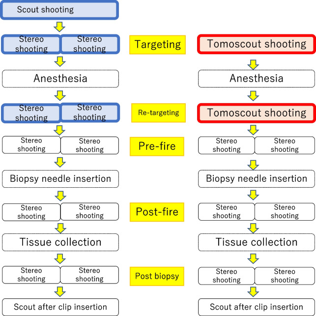
Retrospective review of 131 mammography-guided VABs at our institution were performed. All of the targets were calcification lesion suspicious for cancer. 45 consecutive stereotactic vacuum-assisted biopsies (ST-VABs) and 86 consecutive DBT-VABs were compared. Written informed consent was obtained. Tissue sampling methods and materials were the same with both systems. Student's t-test was used to compare procedure time and the Fisher's exact test was used to compare success rate, complications, and histopathologic findings for the 2 methods.
The tissue sampling success rate was 95.6% for ST-VAB (43/45) and 97.7% (84/86) for DBT-VAB. Time for positioning (10.6 ± 6.4 vs. 6.7 ± 5.3 min), time for biopsy (33.4 ± 13.1 vs. 22.5 ± 13.1 min), and overall procedure time (66.6 ± 16.6 min vs. 54.5 ± 13.0 min) were substantially shorter with DBT-VAB (P < 0.0001). There were no differences in the distribution of pathological findings between the 2 groups.
Depth information and stable visibility of the target provided by DBT images led to quick decisions about target coordinates and improved the clinical performance of microcalcification biopsies.
本研究旨在评估数字乳腺断层合成引导真空辅助活检(DBT-VAB)在乳腺微钙化病变中的临床性能。
对我院 131 例乳腺 X 线摄影引导真空辅助活检(VAB)进行回顾性分析。所有靶标均为可疑钙化的癌性病变。对比了 45 例连续立体定向真空辅助活检(ST-VAB)和 86 例连续 DBT-VAB。所有患者均签署了知情同意书。两种系统的组织取样方法和材料相同。采用 Student's t 检验比较两种方法的手术时间,采用 Fisher 确切概率法比较成功率、并发症和组织病理学结果。
ST-VAB 的组织取样成功率为 95.6%(43/45),DBT-VAB 的成功率为 97.7%(84/86)。定位时间(10.6 ± 6.4 比 6.7 ± 5.3 分钟)、活检时间(33.4 ± 13.1 比 22.5 ± 13.1 分钟)和总手术时间(66.6 ± 16.6 比 54.5 ± 13.0 分钟)在 DBT-VAB 中显著缩短(P < 0.0001)。两组的组织学发现分布无差异。
DBT 图像提供的目标深度信息和稳定的可视性使目标坐标的决策更快,提高了微钙化病变活检的临床性能。