• 文献检索
  • 文档翻译
  • 深度研究
  • 学术资讯
  • Suppr Zotero 插件Zotero 插件
  • 邀请有礼
  • 套餐&价格
  • 历史记录
应用&插件
Suppr Zotero 插件Zotero 插件浏览器插件Mac 客户端Windows 客户端微信小程序
定价
高级版会员购买积分包购买API积分包
服务
文献检索文档翻译深度研究API 文档MCP 服务
关于我们
关于 Suppr公司介绍联系我们用户协议隐私条款
关注我们

Suppr 超能文献

核心技术专利:CN118964589B侵权必究
粤ICP备2023148730 号-1Suppr @ 2026

文献检索

告别复杂PubMed语法,用中文像聊天一样搜索,搜遍4000万医学文献。AI智能推荐,让科研检索更轻松。

立即免费搜索

文件翻译

保留排版,准确专业,支持PDF/Word/PPT等文件格式,支持 12+语言互译。

免费翻译文档

深度研究

AI帮你快速写综述,25分钟生成高质量综述,智能提取关键信息,辅助科研写作。

立即免费体验

使用高频超声和剪切波弹性成像技术对多发性肌炎和皮肌炎患者的肌肉特性进行定量评估。

Quantitative assessment of muscle properties in polymyositis and dermatomyositis using high-frequency ultrasound and shear wave elastography.

作者信息

Li Min, Guo Ruiqian, Tang Xinyi, Huang Songya, Qiu Li

机构信息

Department of Medical Ultrasound, West China Hospital of Sichuan University, Chengdu, China.

出版信息

Quant Imaging Med Surg. 2023 Jan 1;13(1):428-440. doi: 10.21037/qims-22-423. Epub 2022 Nov 22.

DOI:10.21037/qims-22-423
PMID:36620135
原文链接:https://pmc.ncbi.nlm.nih.gov/articles/PMC9816716/
Abstract

BACKGROUND

Polymyositis (PM) and dermatomyositis (DM) are two common types of idiopathic inflammatory myopathy and can lead to a poor prognosis and quality of life. We designed this cross-sectional study to investigate the abilities of high-frequency ultrasound (HFUS) and shear wave elastography (SWE) to assess muscle properties in patients with PM and DM and to distinguish healthy muscles from diseased muscles with PM and DM.

METHODS

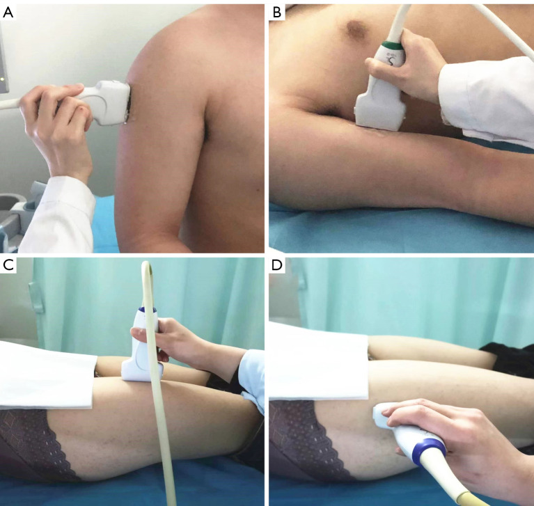
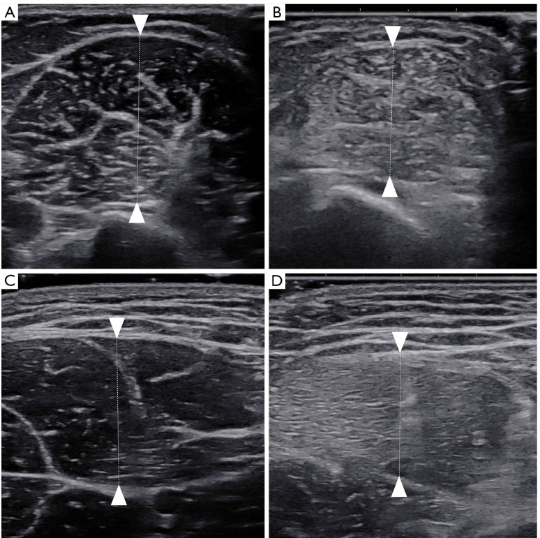
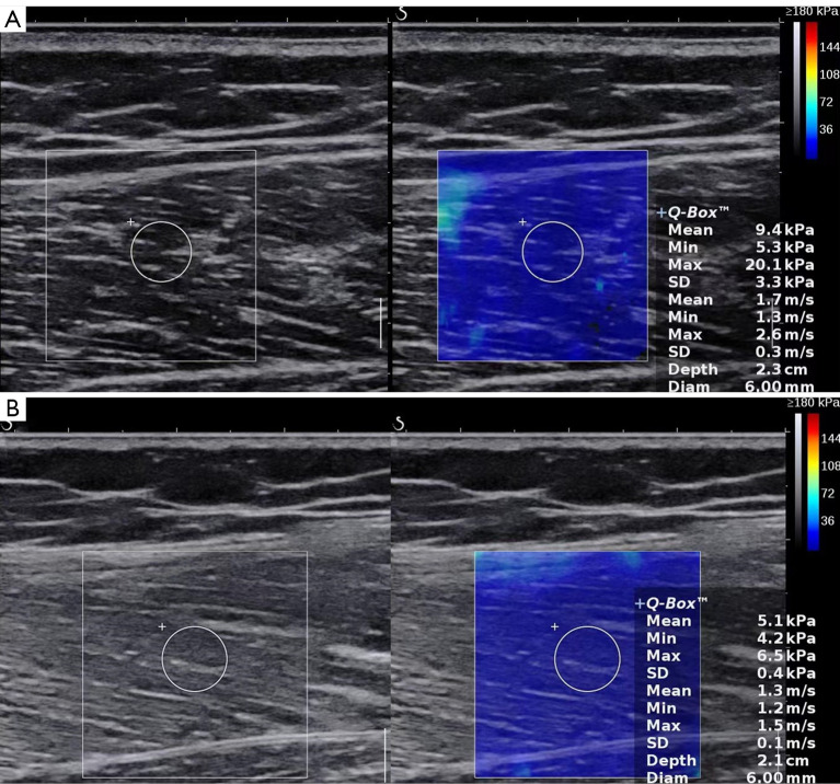
A total of 60 patients (26 PM cases and 34 DM cases) and 65 matched healthy volunteers were continuously included in the case and control groups, respectively. For the bilateral deltoid, biceps brachii, rectus femoris, and vastus lateralis, the muscle thickness, echo intensity, and longitudinal shear wave velocity (SWV) of all participants were measured using HFUS and SWE. The intra- and interobserver reliability of SWV measurements of patients with PM and DM and the receiver operating characteristic curve for HFUS and SWE for PM and DM were analyzed.

RESULTS

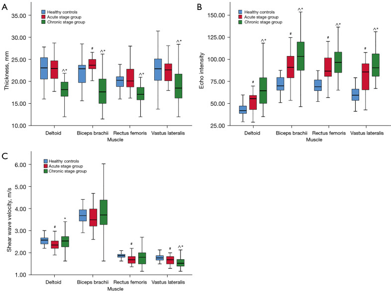
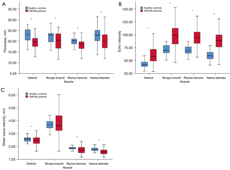
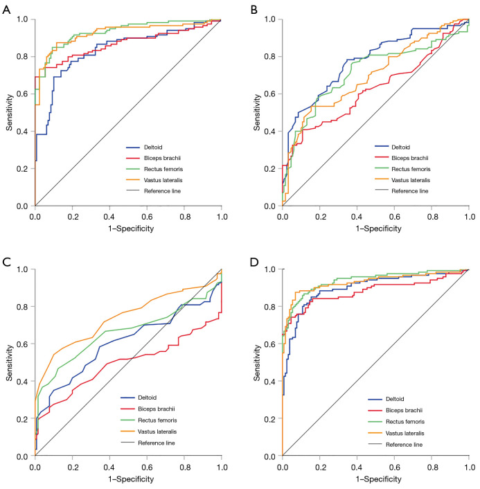
Patients with PM and DM had significantly decreased muscle thickness and increased muscle echo intensity compared to healthy controls (P<0.001). The patients' and healthy participants' deltoid, biceps brachii, rectus femoris, and vastus lateralis thickness was 19.75 and 23.00 mm, 20.45 and 22.80 mm, 18.40 and 20.20 mm, and 20.00 and 22.80 mm, respectively. Except for the biceps brachii, the mean SWV in the longitudinal orientation in patients with PM and DM significantly decreased (P<0.01). The mean SWV of the patients' and healthy participants' deltoid, rectus femoris, and vastus lateralis was 2.47 and 2.57 m/s, 1.73 and 1.87 m/s, and 1.57 and 1.77 m/s, respectively. Excellent intra- and interobserver reliability of SWV measurements on the deltoid and rectus femoris of PM and DM patients were found (intraclass correlation coefficient >0.95; P<0.001). The diagnostic performance of echo intensity in lower-extremity proximal muscles for PM and DM was excellent [area under the curve (AUC) >0.9]. The thickness of most muscles displayed moderate diagnostic performance (the AUC ranged from 0.700 to 0.775). The SWV of the vastus lateralis showed a stable performance (AUC =0.741). The combined diagnostic performance of echo intensity and thickness and the combined diagnostic performance of the 3 indicators were relatively high (the AUC ranged from 0.871 to 0.936 and from 0.898 to 0.938, respectively). Muscle thickness and echo intensity showed statistical differences in different disease stages of PM and DM (P'<0.01).

CONCLUSIONS

HFUS and SWE may serve as imaging biomarkers for the diagnosis of PM and DM by detecting abnormal muscle thinning, enhanced muscle echo intensity, and reduced muscle SWV.

摘要

背景

多发性肌炎(PM)和皮肌炎(DM)是特发性炎性肌病的两种常见类型,可导致不良预后和生活质量下降。我们设计了这项横断面研究,以调查高频超声(HFUS)和剪切波弹性成像(SWE)评估PM和DM患者肌肉特性以及区分健康肌肉与PM和DM患病肌肉的能力。

方法

病例组和对照组分别连续纳入60例患者(26例PM患者和34例DM患者)和65名匹配的健康志愿者。对于双侧三角肌、肱二头肌、股直肌和股外侧肌,使用HFUS和SWE测量所有参与者的肌肉厚度、回声强度和纵向剪切波速度(SWV)。分析了PM和DM患者SWV测量的观察者内和观察者间可靠性以及HFUS和SWE对PM和DM的受试者工作特征曲线。

结果

与健康对照组相比,PM和DM患者的肌肉厚度显著降低,肌肉回声强度增加(P<0.001)。患者和健康参与者的三角肌、肱二头肌、股直肌和股外侧肌厚度分别为19.75和23.00mm、20.45和22.80mm、18.40和20.20mm以及20.00和22.80mm。除肱二头肌外,PM和DM患者纵向方向的平均SWV显著降低(P<0.01)。患者和健康参与者三角肌、股直肌和股外侧肌的平均SWV分别为2.47和2.57m/s、1.73和1.87m/s以及1.57和1.77m/s。发现PM和DM患者三角肌和股直肌SWV测量具有出色的观察者内和观察者间可靠性(组内相关系数>0.95;P<0.001)。下肢近端肌肉回声强度对PM和DM的诊断性能优异[曲线下面积(AUC)>0.9]。大多数肌肉的厚度显示出中等诊断性能(AUC范围为0.700至0.775)。股外侧肌的SWV表现稳定(AUC =0.741)。回声强度和厚度的联合诊断性能以及三个指标的联合诊断性能相对较高(AUC分别范围为0.871至0.936和0.898至0.938)。肌肉厚度和回声强度在PM和DM的不同疾病阶段存在统计学差异(P'<0.01)。

结论

HFUS和SWE可通过检测异常的肌肉变薄、增强的肌肉回声强度和降低的肌肉SWV作为诊断PM和DM的影像学生物标志物。

https://cdn.ncbi.nlm.nih.gov/pmc/blobs/dd0a/9816716/b4662c2fbc20/qims-13-01-428-f6.jpg
https://cdn.ncbi.nlm.nih.gov/pmc/blobs/dd0a/9816716/e044600482d3/qims-13-01-428-f1.jpg
https://cdn.ncbi.nlm.nih.gov/pmc/blobs/dd0a/9816716/8dfb1c14de76/qims-13-01-428-f2.jpg
https://cdn.ncbi.nlm.nih.gov/pmc/blobs/dd0a/9816716/f84822945c18/qims-13-01-428-f3.jpg
https://cdn.ncbi.nlm.nih.gov/pmc/blobs/dd0a/9816716/f784c38e942e/qims-13-01-428-f4.jpg
https://cdn.ncbi.nlm.nih.gov/pmc/blobs/dd0a/9816716/1c46c4fc2961/qims-13-01-428-f5.jpg
https://cdn.ncbi.nlm.nih.gov/pmc/blobs/dd0a/9816716/b4662c2fbc20/qims-13-01-428-f6.jpg

相似文献

1
Quantitative assessment of muscle properties in polymyositis and dermatomyositis using high-frequency ultrasound and shear wave elastography.使用高频超声和剪切波弹性成像技术对多发性肌炎和皮肌炎患者的肌肉特性进行定量评估。
Quant Imaging Med Surg. 2023 Jan 1;13(1):428-440. doi: 10.21037/qims-22-423. Epub 2022 Nov 22.
2
Application of conventional ultrasound coupled with shear wave elastography in the assessment of muscle strength in patients with type 2 diabetes.传统超声联合剪切波弹性成像在2型糖尿病患者肌肉力量评估中的应用
Quant Imaging Med Surg. 2024 Feb 1;14(2):1716-1728. doi: 10.21037/qims-23-1152. Epub 2024 Jan 23.
3
Muscle shear wave elastography in idiopathic inflammatory myopathies: a case-control study with MRI correlation.特发性炎性肌病的肌肉剪切波弹性成像:与 MRI 相关性的病例对照研究。
Skeletal Radiol. 2019 Aug;48(8):1209-1219. doi: 10.1007/s00256-019-03175-3. Epub 2019 Feb 27.
4
Increased fascial thickness of the deltoid muscle in dermatomyositis and polymyositis: An ultrasound study.皮肌炎和多肌炎中三角肌筋膜厚度增加:一项超声研究。
Muscle Nerve. 2015 Oct;52(4):534-9. doi: 10.1002/mus.24595. Epub 2015 Aug 20.
5
The effect of unit, depth, and probe load on the reliability of muscle shear wave elastography: Variables affecting reliability of SWE.单位、深度和探头负荷对肌肉剪切波弹性成像可靠性的影响:影响剪切波弹性成像可靠性的变量
J Clin Ultrasound. 2018 Feb;46(2):108-115. doi: 10.1002/jcu.22534. Epub 2017 Oct 9.
6
Application of ultrasound elastography in the evaluation of muscle strength in a healthy population.超声弹性成像在健康人群肌肉力量评估中的应用。
Quant Imaging Med Surg. 2020 Oct;10(10):1961-1972. doi: 10.21037/qims-20-439.
7
Inter-operator and inter-device reproducibility of shear wave elastography in healthy muscle tissues.健康肌肉组织中剪切波弹性成像的操作者间和设备间可重复性。
J Appl Clin Med Phys. 2022 Sep;23(9):e13717. doi: 10.1002/acm2.13717. Epub 2022 Jul 6.
8
Ultrasound shear wave elastography in assessment of skeletal muscle stiffness in senior volunteers.超声剪切波弹性成像评估老年志愿者骨骼肌硬度。
Clin Imaging. 2019 Nov-Dec;58:22-26. doi: 10.1016/j.clinimag.2019.06.006. Epub 2019 Jun 14.
9
Diagnostic test accuracy of ultrasound for sarcopenia diagnosis: A systematic review and meta-analysis.超声诊断肌肉减少症的准确性:系统评价和荟萃分析。
J Cachexia Sarcopenia Muscle. 2023 Feb;14(1):57-70. doi: 10.1002/jcsm.13149. Epub 2022 Dec 13.
10
Ultrasound Shear Wave Elastography to Assess Osteopathic Manipulative Treatment on the Iliocostalis Lumborum Muscle: A Feasibility Study.超声剪切波弹性成像评估整脊手法治疗竖脊肌的可行性研究。
J Ultrasound Med. 2020 Jan;39(1):157-164. doi: 10.1002/jum.15092. Epub 2019 Jul 4.

引用本文的文献

1
Sarcopenia in Rheumatic Diseases: A Hidden Issue of Concern.风湿性疾病中的肌肉减少症:一个潜藏的关注点。
Diseases. 2025 Apr 26;13(5):134. doi: 10.3390/diseases13050134.
2
Advancing insights: a bibliometric analysis of evolutionary patterns and research frontiers in ultrasound-derived quantitative assessment of skeletal muscle.前沿洞察:超声衍生的骨骼肌定量评估的进化模式与研究前沿的文献计量分析
Quant Imaging Med Surg. 2025 Mar 3;15(3):1912-1926. doi: 10.21037/qims-24-1607. Epub 2025 Feb 26.
3
The value of quantitative muscle ultrasound in monitoring muscle and fascia inflammatory activity in juvenile dermatomyositis patients.

本文引用的文献

1
The Utility of Shear-Wave Elastography in the Evaluation of Myositis.剪切波弹性成像在肌炎评估中的应用。
Ultrasound Med Biol. 2021 Aug;47(8):2176-2185. doi: 10.1016/j.ultrasmedbio.2021.04.010. Epub 2021 May 22.
2
Conventional ultrasound and elastography as imaging outcome tools in autoimmune myositis: A systematic review by the OMERACT ultrasound group.常规超声和弹性成像作为自身免疫性肌炎的影像学结局工具:OMERACT 超声组的系统评价。
Semin Arthritis Rheum. 2021 Jun;51(3):661-676. doi: 10.1016/j.semarthrit.2020.11.001. Epub 2020 Nov 21.
3
Muscle shear wave elastography in idiopathic inflammatory myopathies: a case-control study with MRI correlation.
定量肌肉超声在监测青少年皮肌炎患者肌肉和筋膜炎症活动中的价值。
Quant Imaging Med Surg. 2024 Dec 5;14(12):8601-8613. doi: 10.21037/qims-24-1035. Epub 2024 Nov 24.
4
Imaging mimickers of MSK infection.肌肉骨骼系统感染的影像学表现。
Skeletal Radiol. 2024 Oct;53(10):2227-2246. doi: 10.1007/s00256-024-04763-8. Epub 2024 Aug 16.
5
Muscle ultrasound in myopathies.肌肉超声在肌病中的应用。
Curr Opin Neurol. 2024 Oct 1;37(5):549-557. doi: 10.1097/WCO.0000000000001306. Epub 2024 Jul 25.
6
A Case-Control Study of 40 Patients with Piriformis Muscle Syndrome to Evaluate Diagnostic Findings Using Two-Dimensional Ultrasound and Shear Wave Elastography.一项针对 40 例梨状肌综合征患者的病例对照研究,旨在使用二维超声和剪切波弹性成像评估诊断结果。
Med Sci Monit. 2023 Jun 14;29:e940214. doi: 10.12659/MSM.940214.
特发性炎性肌病的肌肉剪切波弹性成像:与 MRI 相关性的病例对照研究。
Skeletal Radiol. 2019 Aug;48(8):1209-1219. doi: 10.1007/s00256-019-03175-3. Epub 2019 Feb 27.
4
Factors associated with masseter muscle quality assessed from ultrasonography in community-dwelling elderly individuals: A cross-sectional study.社区居住老年人超声评估的咬肌肌肉质量的相关因素:一项横断面研究。
Arch Gerontol Geriatr. 2019 May-Jun;82:128-132. doi: 10.1016/j.archger.2019.02.003. Epub 2019 Feb 12.
5
Imaging in the diagnosis of idiopathic inflammatory myopathies; indications and utility.特发性炎性肌病的影像学诊断;适应证和应用。
Expert Rev Neurother. 2019 Feb;19(2):173-184. doi: 10.1080/14737175.2019.1572507. Epub 2019 Feb 7.
6
Clinical indications for musculoskeletal ultrasound updated in 2017 by European Society of Musculoskeletal Radiology (ESSR) consensus.2017 年,欧洲肌肉骨骼放射学会(ESSR)通过共识更新了肌肉骨骼超声的临床适应证。
Eur Radiol. 2018 Dec;28(12):5338-5351. doi: 10.1007/s00330-018-5474-3. Epub 2018 Jun 6.
7
Pattern of muscle involvement in inclusion body myositis: a sonographic study.包涵体肌炎的肌肉受累模式:超声研究。
Clin Exp Rheumatol. 2018 Nov-Dec;36(6):996-1002. Epub 2018 May 8.
8
Muscle Shear Wave Elastography in Inclusion Body Myositis: Feasibility, Reliability and Relationships with Muscle Impairments.包涵体肌炎中的肌肉剪切波弹性成像:可行性、可靠性及与肌肉损伤的关系
Ultrasound Med Biol. 2018 Jul;44(7):1423-1432. doi: 10.1016/j.ultrasmedbio.2018.03.026. Epub 2018 Apr 26.
9
The effect of unit, depth, and probe load on the reliability of muscle shear wave elastography: Variables affecting reliability of SWE.单位、深度和探头负荷对肌肉剪切波弹性成像可靠性的影响:影响剪切波弹性成像可靠性的变量
J Clin Ultrasound. 2018 Feb;46(2):108-115. doi: 10.1002/jcu.22534. Epub 2017 Oct 9.
10
An efficacy analysis of whole-body magnetic resonance imaging in the diagnosis and follow-up of polymyositis and dermatomyositis.全身磁共振成像在多发性肌炎和皮肌炎诊断及随访中的疗效分析
PLoS One. 2017 Jul 17;12(7):e0181069. doi: 10.1371/journal.pone.0181069. eCollection 2017.