From the Department of Radiology, Research Institute for Convergence of Biomedical Science and Technology, Pusan National University Yangsan Hospital, Pusan National University School of Medicine, Yangsan, Korea (Y.J.J.); Division of Infectious Diseases, Department of Internal Medicine (Y.M.W., S.H.K.) and Department of Radiology (K.S.L.), Samsung Changwon Hospital, Sungkyunkwan University School of Medicine (SKKU-SOM), Changwon 51353, Korea; Department of Electrical and Computer Engineering, Sungkyunkwan University, Suwon, Korea (H.P.); Center for Neuroscience Imaging Research, Institute for Basic Science, Suwon, Korea (H.P.); and Department of Radiology, Chonnam National University Hospital, Gwangju, Korea (J.E.L.).
Radiology. 2023 Feb;306(2):e222462. doi: 10.1148/radiol.222462. Epub 2023 Jan 10.
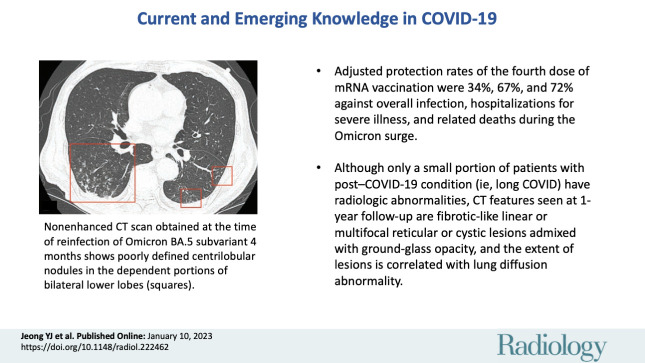
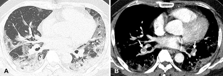
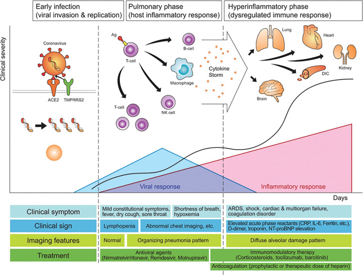
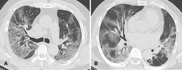
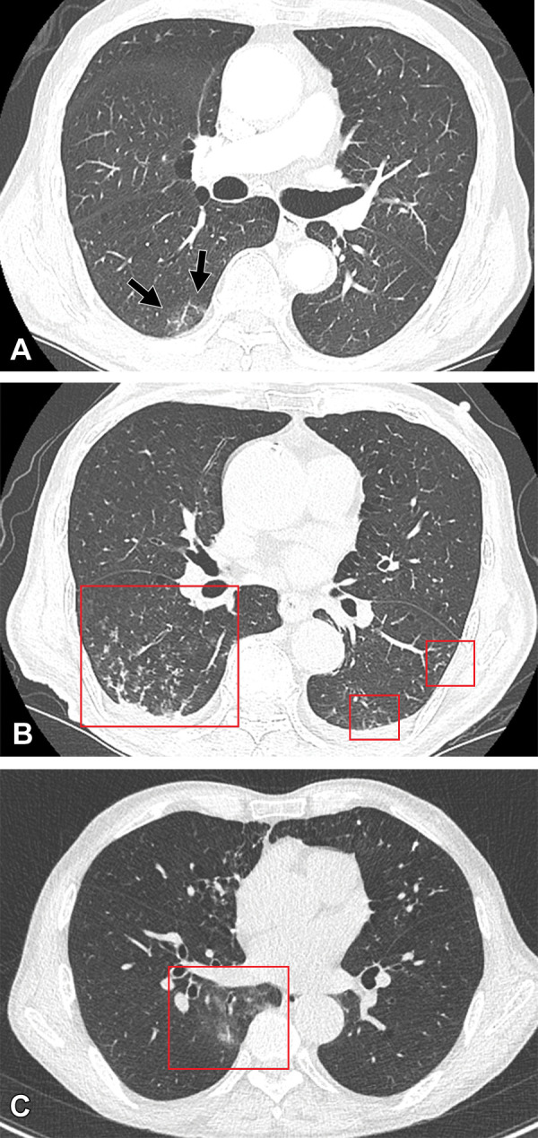
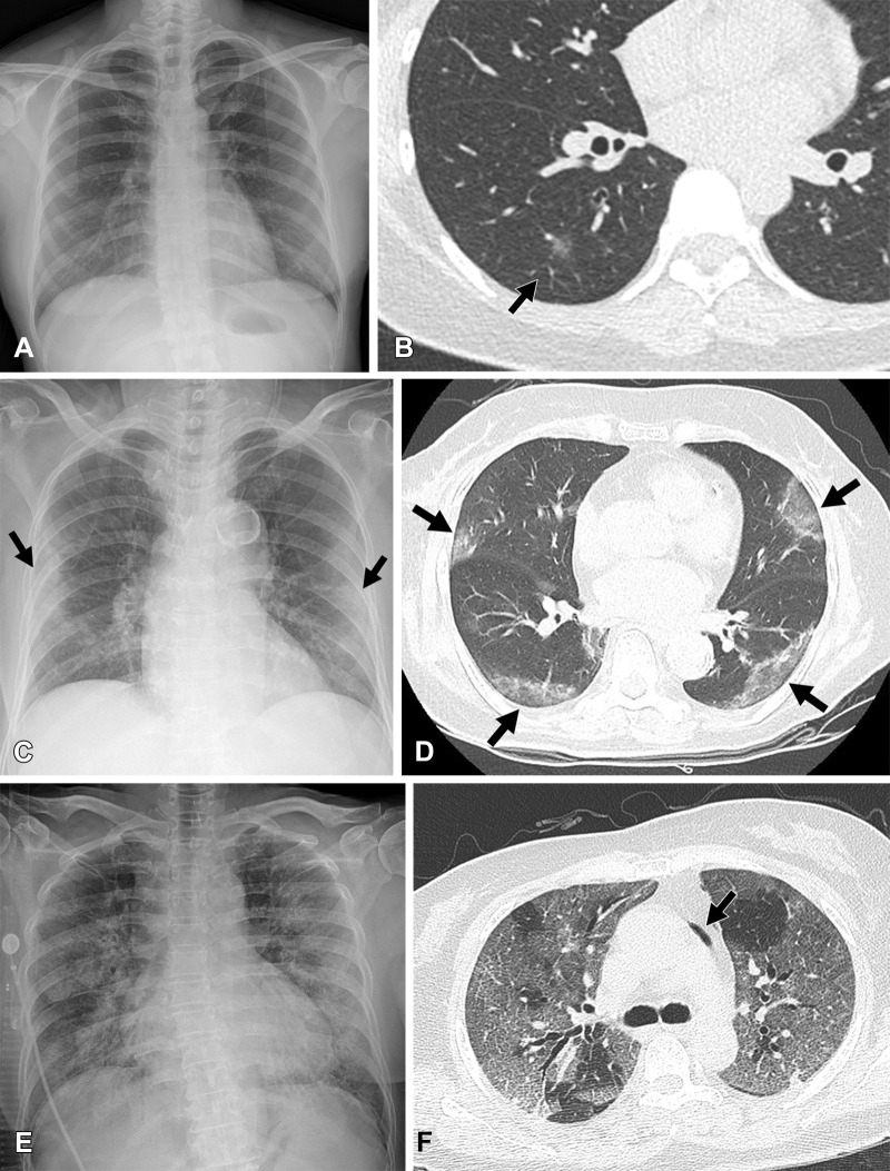
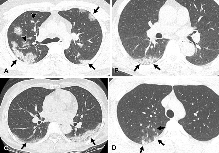
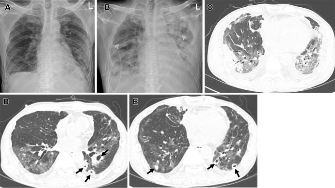
COVID-19 has emerged as a pandemic leading to a global public health crisis of unprecedented morbidity. A comprehensive insight into the imaging of COVID-19 has enabled early diagnosis, stratification of disease severity, and identification of potential sequelae. The evolution of COVID-19 can be divided into early infectious, pulmonary, and hyperinflammatory phases. Clinical features, imaging features, and management are different among the three phases. In the early stage, peripheral ground-glass opacities are predominant CT findings, and therapy directly targeting SARS-CoV-2 is effective. In the later stage, organizing pneumonia or diffuse alveolar damage pattern are predominant CT findings and anti-inflammatory therapies are more beneficial. The risk of severe disease or hospitalization is lower in breakthrough or Omicron variant infection compared with nonimmunized or Delta variant infections. The protection rates of the fourth dose of mRNA vaccination were 34% and 67% against overall infection and hospitalizations for severe illness, respectively. After acute COVID-19 pneumonia, most residual CT abnormalities gradually decreased in extent, but they may remain as linear or multifocal reticular or cystic lesions. Advanced insights into the pathophysiologic and imaging features of COVID-19 along with vaccine benefits have improved patient care, but emerging knowledge of post-COVID-19 condition, or long COVID, also presents radiology with new challenges.
新型冠状病毒肺炎(COVID-19)已成为一种大流行疾病,引发了前所未有的全球公共卫生危机。对 COVID-19 影像学的全面了解使早期诊断、疾病严重程度分层和潜在后遗症的识别成为可能。COVID-19 的演变可分为早期感染、肺部和超炎症期。这三个阶段的临床特征、影像学特征和治疗方法都有所不同。在早期,外周磨玻璃影是 CT 的主要发现,直接针对 SARS-CoV-2 的治疗是有效的。在后期,机化性肺炎或弥漫性肺泡损伤模式是 CT 的主要发现,抗炎治疗更为有益。与非免疫或德尔塔变异感染相比,突破性或奥密克戎变异感染发生重症疾病或住院治疗的风险较低。mRNA 疫苗第四剂的保护率分别为 34%和 67%,可预防总体感染和因重症疾病住院治疗。急性 COVID-19 肺炎后,大多数残留的 CT 异常逐渐减少,但可能仍然存在线性或多灶性网状或囊性病变。对 COVID-19 的病理生理和影像学特征以及疫苗益处的深入了解改善了患者的治疗,但 COVID-19 后状况(即长新冠)的新知识也给放射学带来了新的挑战。