• 文献检索
  • 文档翻译
  • 深度研究
  • 学术资讯
  • Suppr Zotero 插件Zotero 插件
  • 邀请有礼
  • 套餐&价格
  • 历史记录
应用&插件
Suppr Zotero 插件Zotero 插件浏览器插件Mac 客户端Windows 客户端微信小程序
定价
高级版会员购买积分包购买API积分包
服务
文献检索文档翻译深度研究API 文档MCP 服务
关于我们
关于 Suppr公司介绍联系我们用户协议隐私条款
关注我们

Suppr 超能文献

核心技术专利:CN118964589B侵权必究
粤ICP备2023148730 号-1Suppr @ 2026

文献检索

告别复杂PubMed语法,用中文像聊天一样搜索,搜遍4000万医学文献。AI智能推荐,让科研检索更轻松。

立即免费搜索

文件翻译

保留排版,准确专业,支持PDF/Word/PPT等文件格式,支持 12+语言互译。

免费翻译文档

深度研究

AI帮你快速写综述,25分钟生成高质量综述,智能提取关键信息,辅助科研写作。

立即免费体验

基于多模态超声的颈动脉斑块风险生物标志物可预测缺血性卒中和 TIA 患者的不良功能结局。

Multimodal ultrasound-based carotid plaque risk biomarkers predict poor functional outcome in patients with ischemic stroke or TIA.

机构信息

Department of Ultrasound, Beijing Tiantan Hospital, Capital Medical University, No.119 South Fourth Ring West Road, Fengtai District, Beijing, 100160, China.

Guang'anmen Hospital, Chinese Academy of traditional Chinese Medicine, Beijing, China.

出版信息

BMC Neurol. 2023 Jan 12;23(1):13. doi: 10.1186/s12883-023-03052-6.

DOI:10.1186/s12883-023-03052-6
PMID:36631804
原文链接:https://pmc.ncbi.nlm.nih.gov/articles/PMC9835263/
Abstract

BACKGROUND

Carotid vulnerable plaque is an important risk factor for stroke occurrence and recurrence. However, the relationship between risk parameters related to carotid vulnerable plaque (plaque size, echogenicity, intraplaque neovascularization, and plaque stiffness) and neurological outcome after ischemic stroke or TIA is unclear. This study investigates the value of multimodal ultrasound-based carotid plaque risk biomarkers to predict poor short-term functional outcome after ischemic stroke or TIA.

METHODS

This study was a single-center, prospective, continuous, cohort study to observe the occurrence of adverse functional outcomes (mRS 2-6/3-6) 90 days after ischemic stroke or TIA in patients, where the exposure factors in this study were carotid plaque ultrasound risk biomarkers and the risk factors were sex, age, disease history, and medication history. Patients with ischemic stroke or TIA (mRS ≤3) whose ipsilateral internal carotid artery stenosis was ≥50% within 30 days were included. All patients underwent multimodal ultrasound at baseline, including conventional ultrasound, superb microvascular imaging (SMI), and shear wave elastography (SWE). Continuous variables were divided into four groups at interquartile spacing for inclusion in univariate and multifactorial analyses. After completion of a baseline ultrasound, all patients were followed up at 90 days after ultrasound, and patient modified neurological function scores (mRSs) were recorded. Multivariate Cox regression and ROC curves were used to assess the risk factors and predictive power for predicting poor neurological function.

RESULTS

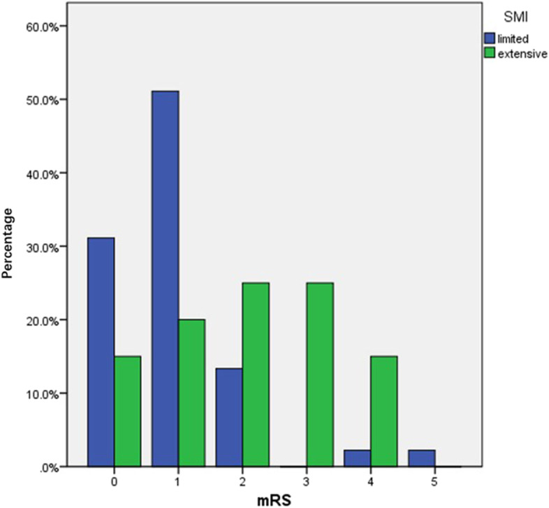
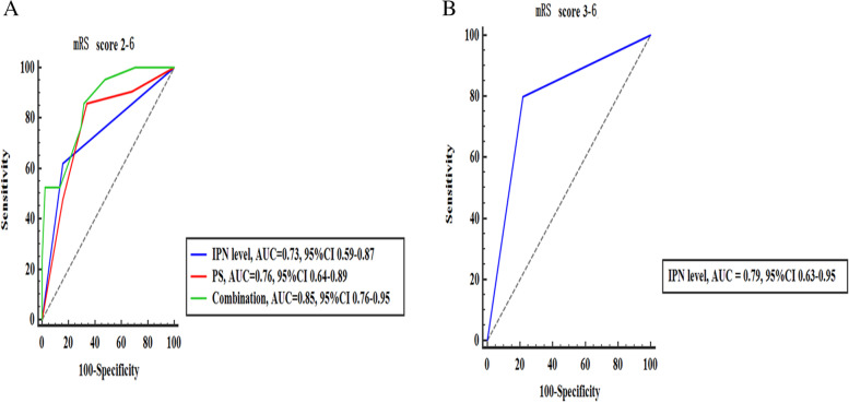
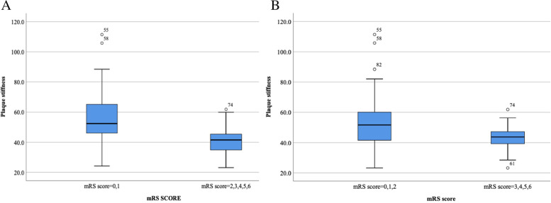
SMI revealed that 20 (30.8%) patients showed extensive neovascularization in the carotid plaque, and 45 (69.2%) patients showed limited neovascularization in the carotid plaque. SWE imaging showed that the mean carotid plaque stiffness was 51.49 ± 18.34 kPa (23.19-111.39 kPa). After a mean follow-up of 90 ± 14 days, a total of 21 (32.3%) patients had a mRS of 2-6, and a total of 10 (15.4%) patients had a mRS of 3-6. Cox regression analysis showed that the level of intraplaque neovascularization and plaque stiffness were independent risk factors for a mRS of 2-6, and the level of intraplaque neovascularization was an independent risk factor for a mRS of 3-6. After correcting for confounders, the HR of intraplaque neovascularization level and plaque stiffness predicting a mRS 2-6 was 3.06 (95% CI 1.05-12.59, P = 0.041) and 0.51 (95% CI 0.31-0.83, P = 0.007), respectively; the HR of intraplaque neovascularization level predicting a mRS 3-6 was 6.11 (95% CI 1.19-31.45, P = 0.031). For ROC curve analysis, the mRSs for intraplaque neovascularization level, plaque stiffness, and combined application to predict 90-day neurological outcome ranged from 2 to 6, with AUCs of 0.73 (95% CI 0.59-0.87), 0.76 (95% CI 0.64-0.89) and 0.85 (95% CI 0.76-0.95), respectively. The mRSs for the intraplaque neovascularization level to predict 90-day neurological outcome ranged from 3 to 6, with AUCs of 0.79 (95% CI 0.63-0.95).

CONCLUSION

Intraplaque neovascularization level and plaque stiffness may be associated with an increased risk of poor short-term functional outcome after stroke in patients with recent anterior circulation ischemic stroke due to carotid atherosclerosis. The combined application of multiple parameters has efficacy in predicting poor short-term functional outcome after stroke.

摘要

背景

颈动脉易损斑块是中风发生和复发的一个重要危险因素。然而,颈动脉易损斑块的风险参数(斑块大小、回声、斑块内新生血管形成和斑块硬度)与缺血性卒中和 TIA 后神经功能结局之间的关系尚不清楚。本研究旨在探讨基于多模态超声的颈动脉斑块风险生物标志物预测颈动脉粥样硬化性缺血性卒中和 TIA 后近期不良短期功能结局的价值。

方法

这是一项单中心、前瞻性、连续、队列研究,观察缺血性卒中和 TIA 后 90 天患者不良功能结局(mRS 2-6/3-6)的发生情况,本研究中的暴露因素为颈动脉斑块超声风险生物标志物,风险因素为性别、年龄、病史和用药史。纳入的患者为同侧颈内动脉狭窄≥50%的缺血性卒中和 TIA(mRS≤3)患者。所有患者均在基线时进行多模态超声检查,包括常规超声、超微血流成像(SMI)和剪切波弹性成像(SWE)。连续变量按四分位间距分为四组,纳入单因素和多因素分析。完成基线超声检查后,所有患者在超声检查后 90 天进行随访,并记录患者改良神经功能评分(mRSs)。多因素 Cox 回归和 ROC 曲线用于评估预测不良神经功能的风险因素和预测能力。

结果

SMI 显示 20 例(30.8%)患者颈动脉斑块内广泛新生血管形成,45 例(69.2%)患者颈动脉斑块内局限新生血管形成。SWE 成像显示颈动脉斑块平均硬度为 51.49±18.34kPa(23.19-111.39kPa)。平均随访 90±14 天后,共有 21 例(32.3%)患者 mRS 为 2-6,共有 10 例(15.4%)患者 mRS 为 3-6。Cox 回归分析显示,斑块内新生血管形成水平和斑块硬度是 mRS 为 2-6 的独立危险因素,斑块内新生血管形成水平是 mRS 为 3-6 的独立危险因素。在纠正混杂因素后,斑块内新生血管形成水平和斑块硬度预测 mRS 2-6 的 HR 分别为 3.06(95%CI 1.05-12.59,P=0.041)和 0.51(95%CI 0.31-0.83,P=0.007);斑块内新生血管形成水平预测 mRS 3-6 的 HR 为 6.11(95%CI 1.19-31.45,P=0.031)。对于 ROC 曲线分析,斑块内新生血管形成水平、斑块硬度和联合应用预测 90 天神经功能结局的 mRS 范围为 2-6,AUC 分别为 0.73(95%CI 0.59-0.87)、0.76(95%CI 0.64-0.89)和 0.85(95%CI 0.76-0.95)。斑块内新生血管形成水平预测 90 天神经功能结局的 mRS 范围为 3-6,AUC 为 0.79(95%CI 0.63-0.95)。

结论

颈动脉粥样硬化性缺血性卒中和 TIA 患者近期,斑块内新生血管形成水平和斑块硬度可能与不良短期功能结局风险增加相关。多个参数的联合应用在预测缺血性卒中和 TIA 后近期不良短期功能结局方面具有一定的预测价值。

https://cdn.ncbi.nlm.nih.gov/pmc/blobs/e697/9835263/bd1777fb03df/12883_2023_3052_Fig3_HTML.jpg
https://cdn.ncbi.nlm.nih.gov/pmc/blobs/e697/9835263/62b1f27dc5aa/12883_2023_3052_Fig1_HTML.jpg
https://cdn.ncbi.nlm.nih.gov/pmc/blobs/e697/9835263/2093f7b9078d/12883_2023_3052_Fig2_HTML.jpg
https://cdn.ncbi.nlm.nih.gov/pmc/blobs/e697/9835263/bd1777fb03df/12883_2023_3052_Fig3_HTML.jpg
https://cdn.ncbi.nlm.nih.gov/pmc/blobs/e697/9835263/62b1f27dc5aa/12883_2023_3052_Fig1_HTML.jpg
https://cdn.ncbi.nlm.nih.gov/pmc/blobs/e697/9835263/2093f7b9078d/12883_2023_3052_Fig2_HTML.jpg
https://cdn.ncbi.nlm.nih.gov/pmc/blobs/e697/9835263/bd1777fb03df/12883_2023_3052_Fig3_HTML.jpg

相似文献

1
Multimodal ultrasound-based carotid plaque risk biomarkers predict poor functional outcome in patients with ischemic stroke or TIA.基于多模态超声的颈动脉斑块风险生物标志物可预测缺血性卒中和 TIA 患者的不良功能结局。
BMC Neurol. 2023 Jan 12;23(1):13. doi: 10.1186/s12883-023-03052-6.
2
Advanced ultrasound methods in assessment of carotid plaque instability: a prospective multimodal study.颈动脉斑块不稳定性评估的高级超声方法:一项前瞻性多模态研究。
BMC Neurol. 2020 Jan 29;20(1):39. doi: 10.1186/s12883-020-1620-z.
3
Ultrasound Characteristics of Symptomatic Carotid Plaques: A Systematic Review and Meta-Analysis.有症状颈动脉斑块的超声特征:一项系统评价与Meta分析
Cerebrovasc Dis. 2015;40(3-4):165-74. doi: 10.1159/000437339. Epub 2015 Aug 13.
4
Carotid Plaque Characteristics Predict Recurrent Ischemic Stroke and TIA: The PARISK (Plaque At RISK) Study.颈动脉斑块特征预测复发性缺血性中风和短暂性脑缺血发作:PARISK(风险斑块)研究。
JACC Cardiovasc Imaging. 2022 Oct;15(10):1715-1726. doi: 10.1016/j.jcmg.2022.04.003. Epub 2022 Jun 15.
5
Intraplaque Microvascular Flow Signal in Superb Microvascular Imaging and Magnetic Resonance Imaging Carotid Plaque Imaging in Patients with Atheromatous Carotid Artery Stenosis.动脉粥样硬化性颈动脉狭窄患者在高分辨率微血管成像和磁共振成像颈动脉斑块成像中的斑块内微血管血流信号
J Stroke Cerebrovasc Dis. 2018 Dec;27(12):3529-3534. doi: 10.1016/j.jstrokecerebrovasdis.2018.08.017. Epub 2018 Sep 7.
6
Extracranial carotid plaque hemorrhage predicts ipsilateral stroke recurrence in patients with carotid atherosclerosis - a study based on high-resolution vessel wall imaging MRI.基于高分辨率血管壁成像 MRI 的研究:颅外颈动脉斑块出血可预测颈动脉粥样硬化患者同侧卒中复发。
BMC Neurol. 2022 Jun 28;22(1):237. doi: 10.1186/s12883-022-02758-3.
7
Complicated Carotid Artery Plaques and Risk of Recurrent Ischemic Stroke or TIA.复杂颈动脉斑块与复发性缺血性卒中和 TIA 风险。
J Am Coll Cardiol. 2022 Jun 7;79(22):2189-2199. doi: 10.1016/j.jacc.2022.03.376. Epub 2022 May 3.
8
Value of Intraplaque Neovascularization on Contrast-Enhanced Ultrasonography in Predicting Ischemic Stroke Recurrence in Patients With Carotid Atherosclerotic Plaque.颈动脉粥样硬化斑块患者斑块内新生血管在对比增强超声中的价值对预测缺血性脑卒中复发的价值。
Korean J Radiol. 2023 Apr;24(4):338-348. doi: 10.3348/kjr.2022.0977. Epub 2023 Feb 16.
9
Advance ultrasound techniques for the assessment of plaque vulnerability in symptomatic and asymptomatic carotid stenosis: a multimodal ultrasound study.用于评估有症状和无症状颈动脉狭窄斑块易损性的先进超声技术:一项多模态超声研究
Cardiovasc Diagn Ther. 2021 Feb;11(1):28-38. doi: 10.21037/cdt-20-876.
10
Association of Progression of Carotid Artery Wall Volume and Recurrent Transient Ischemic Attack or Stroke: A Magnetic Resonance Imaging Study.颈动脉壁体积进展与复发性短暂性脑缺血发作或卒中的相关性:一项磁共振成像研究。
Stroke. 2018 Mar;49(3):614-620. doi: 10.1161/STROKEAHA.117.019422. Epub 2018 Jan 30.

引用本文的文献

1
Association between carotid plaque calcification and clinical outcomes of symptomatic cerebral small vessel disease.颈动脉斑块钙化与有症状脑小血管病临床结局之间的关联
Front Neurol. 2025 Aug 14;16:1628353. doi: 10.3389/fneur.2025.1628353. eCollection 2025.
2
Predictive value of contrast-enhanced ultrasound in assessing the risk of recurrent in ipsilateral anterior circulation ischemic stroke.超声造影在评估同侧前循环缺血性脑卒中复发风险中的预测价值
BMC Med Imaging. 2025 Aug 26;25(1):350. doi: 10.1186/s12880-025-01887-8.
3
Morphological and biomechanical characteristics of carotid plaques with mild stenosis using ultrafast and high-definition ultrasonography.

本文引用的文献

1
Carotid Intraplaque Neovascularization Predicts Ischemic Stroke Recurrence in Patients with Carotid Atherosclerosis.颈动脉斑块内新生血管与颈动脉粥样硬化患者缺血性卒中复发的相关性研究。
Gerontology. 2021;67(2):144-151. doi: 10.1159/000511360. Epub 2021 Feb 12.
2
Stress-associated neurobiological activity is linked with acute plaque instability via enhanced macrophage activity: a prospective serial 18F-FDG-PET/CT imaging assessment.应激相关的神经生物学活性通过增强巨噬细胞活性与急性斑块不稳定相关:一项前瞻性系列 18F-FDG-PET/CT 成像评估。
Eur Heart J. 2021 May 14;42(19):1883-1895. doi: 10.1093/eurheartj/ehaa1095.
3
Inflammatory markers as independent predictors for stroke outcomes.
使用超快高清超声检查法对轻度狭窄颈动脉斑块的形态学和生物力学特征进行研究。
Eur Radiol. 2025 Jul 14. doi: 10.1007/s00330-025-11816-6.
4
Independent predictors of ischemic stroke: a comprehensive scan of the internal carotid and vertebrobasilar systems.缺血性卒中的独立预测因素:颈内动脉和椎基底动脉系统的全面扫描
Quant Imaging Med Surg. 2025 Apr 1;15(4):2827-2838. doi: 10.21037/qims-24-1763. Epub 2025 Mar 14.
5
Napkin-ring sign plaques are associated with cerebral small vessel disease.餐巾环征斑块与脑小血管病相关。
Eur J Med Res. 2025 Mar 21;30(1):190. doi: 10.1186/s40001-025-02371-3.
6
Incremental clinical value of intraplaque neovascularization in predicting recurrent ischemic stroke.斑块内新生血管形成在预测复发性缺血性卒中方面的增量临床价值。
Ann Clin Transl Neurol. 2025 Feb;12(2):291-299. doi: 10.1002/acn3.52255. Epub 2024 Nov 18.
7
Analysis of carotid ultrasound in a high-stroke-risk population.颈动脉超声分析在高卒中风险人群中的应用。
Medicine (Baltimore). 2024 Nov 1;103(44):e40383. doi: 10.1097/MD.0000000000040383.
8
Relationship between maximum carotid plaque area and prognosis of patients with acute ischaemic stroke.颈动脉斑块最大面积与急性缺血性脑卒中患者预后的关系。
Ir J Med Sci. 2024 Oct;193(5):2501-2507. doi: 10.1007/s11845-024-03739-w. Epub 2024 Jun 18.
炎症标志物作为中风结局的独立预测因子。
Brain Behav. 2021 Jan;11(1):e01922. doi: 10.1002/brb3.1922. Epub 2020 Dec 12.
4
Prognostic Metrics Associated with Inflammation and Atherosclerosis Signaling Evaluate the Burden of Adverse Clinical Outcomes in Ischemic Stroke Patients.与炎症和动脉粥样硬化信号相关的预后指标评估缺血性脑卒中患者不良临床结局的负担。
Clin Chem. 2020 Nov 1;66(11):1434-1443. doi: 10.1093/clinchem/hvaa201.
5
Prevalence of High-risk Plaques and Risk of Stroke in Patients With Asymptomatic Carotid Stenosis: A Meta-analysis.无症状性颈动脉狭窄患者高危斑块的患病率和卒中风险:一项荟萃分析。
JAMA Neurol. 2020 Dec 1;77(12):1524-1535. doi: 10.1001/jamaneurol.2020.2658.
6
Vulnerable plaques and patients: state-of-the-art.易损斑块与患者:现状分析。
Eur Heart J. 2020 Aug 14;41(31):2997-3004. doi: 10.1093/eurheartj/ehaa227.
7
Prevalence of Ipsilateral Nonstenotic Carotid Plaques on Computed Tomography Angiography in Embolic Stroke of Undetermined Source.不明来源栓塞性卒中患者计算机断层血管造影术同侧非狭窄性颈动脉斑块的患病率。
Stroke. 2020 Jun;51(6):1743-1749. doi: 10.1161/STROKEAHA.120.029404. Epub 2020 May 7.
8
Plasma myeloperoxidase levels in acute brain ischaemia and high grade carotid stenosis.急性脑缺血和颈动脉高度狭窄患者的血浆髓过氧化物酶水平。
Eur J Neurol. 2020 Aug;27(8):1604-1611. doi: 10.1111/ene.14279. Epub 2020 May 25.
9
Plaque neovascularization detected with contrast-enhanced ultrasound predicts ischaemic stroke recurrence in patients with carotid atherosclerosis.颈动脉粥样硬化患者经超声造影检测到的斑块新生血管化可预测缺血性卒中复发。
Eur J Neurol. 2020 May;27(5):809-816. doi: 10.1111/ene.14157. Epub 2020 Feb 16.
10
Prediction of Stroke Risk by Detection of Hemorrhage in Carotid Plaques: Meta-Analysis of Individual Patient Data.基于颈动脉斑块出血检测预测卒中风险:个体患者数据分析的荟萃分析。
JACC Cardiovasc Imaging. 2020 Feb;13(2 Pt 1):395-406. doi: 10.1016/j.jcmg.2019.03.028. Epub 2019 Jun 12.