Lowry Ellen, Coughlan Gillian, Morrissey Sol, Jeffs Stephen, Hornberger Michael
Norwich Medical School, University of East Anglia, Norwich NR4 7TJ, United Kingdom.
Harvard Medical School, Massachusetts General Hospital, United States.
Cereb Circ Cogn Behav. 2022 Dec 16;4:100155. doi: 10.1016/j.cccb.2022.100155. eCollection 2023.
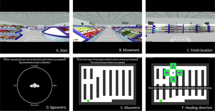
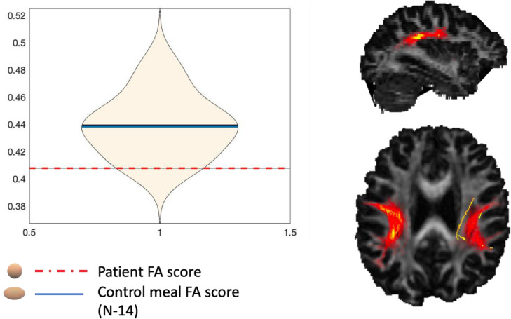
Vascular cognitive impairment (VCI) is the second most prevalent form of dementia, but little is known about the early cognitive and neuroimaging markers. Spatial navigation deficits are an emerging marker for Alzheimer's disease (AD), yet less is known about spatial orientation deficits sensitive to VCI. This case report follows up on the first VCI patient identified to have an egocentric orientation deficit. The study aimed to examine the patient's spatial deficits three years on and gain insights from the addition of the patient's MRI brain scan. A battery of spatial navigation tasks were administered following neuropsychological assessment. Results continue to show spatial orientation deficits. Critically, these changes appear stable and are sensitive to novel spatial tests. Whereas conventional screening tools demonstrate patient recovery. MRI DTI analysis indicates a non-significant trend towards loss of structural integrity to the posterior tracts of the longitudinal superior fasciculus (SLF), while the medial temporal lobe, typically implicated in spatial navigation, is unaffected. This finding potentially reflects reduced network connectivity in posterior to anterior white matter tracts co-existing with spatial orientation deficits. Findings have clinical utility and show spatial orientation as a potential sensitive cognitive marker for VCI.
血管性认知障碍(VCI)是第二常见的痴呆形式,但对于早期认知和神经影像标志物知之甚少。空间导航缺陷是阿尔茨海默病(AD)的一种新出现的标志物,但对于对VCI敏感的空间定向缺陷了解较少。本病例报告追踪了首例被确定存在自我中心定向缺陷的VCI患者。该研究旨在三年后检查该患者的空间缺陷,并通过增加患者的脑部MRI扫描获得见解。在神经心理学评估后进行了一系列空间导航任务。结果继续显示存在空间定向缺陷。关键的是,这些变化似乎是稳定的,并且对新颖的空间测试敏感。而传统的筛查工具显示患者已康复。MRI弥散张量成像(DTI)分析表明,纵向额上束(SLF)后束的结构完整性丧失呈不显著趋势,而通常与空间导航有关的内侧颞叶未受影响。这一发现可能反映了后向前白质束的网络连接减少,同时存在空间定向缺陷。研究结果具有临床实用性,并表明空间定向是VCI的一个潜在敏感认知标志物。