Department of Ophthalmology, Gifu University Graduate School of Medicine, Gifu, Japan.
Ophthalmology, Ogaki Municipal Hospital, Ogaki, Japan.
PLoS One. 2023 Jan 12;18(1):e0278234. doi: 10.1371/journal.pone.0278234. eCollection 2023.
To determine whether multifocal electroretinograms (mfERGs) recorded with natural pupils and skin electrodes can be used to determine the stage of open angle glaucoma (OAG).
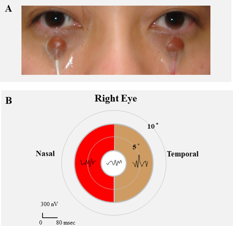
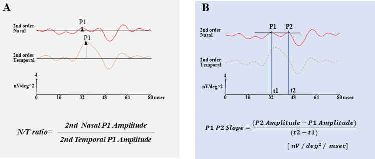
Two hundred eighteen eyes of 132 OAG patients and 62 eyes of 62 normal subjects whose best-corrected visual acuity (BCVA) was 0.1 logarithm of the minimum angle of resolution (logMAR) units (20/25) or less were studied. The mean deviations (MDs) obtained by Humphrey Visual Field Analyzer (HFA), optical coherence tomographic (OCT) images, and mfERGs were analyzed. The glaucoma was classified into 4 stages: preperimetric glaucoma (PPG), early stage, moderate stage, and advanced stage glaucoma. The parameters of the mfERGs examined were the amplitudes of the two positive peaks (P1, P2) of the second order kernels in the nasal and temporal fields within the central 15° diameter.
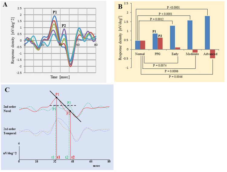
The mean age of all participants (patients and normals) was 63.8 ± 10.8 years. With the progression of glaucoma, the amplitudes of P1 in the nasal hemifield increased and the amplitudes of P2 decreased. The nasal to temporal ratio (N/T ratio) of the P1 amplitudes and the negative slope of the line between P1 and P2 (P1P2 Slope) in the nasal field were larger at each glaucoma stage except at the PPG stage. Both the N/T amplitude ratio and P1P2 Slope were weakly but significantly correlated with the MD (r = -0.3139, P<0.0001; r = 0.4501, P<0.0001, respectively), and the OCT parameters (all P<0.0001) except the outer layer thickness.
Our findings indicate that the amplitudes of P1 and P2 of the second order kernel of the mfERGs in the nasal field of the center region can be good markers for the stages of glaucoma.
确定使用自然瞳孔和皮肤电极记录的多焦视网膜电图(mfERG)是否可用于确定开角型青光眼(OAG)的阶段。
研究了 132 例 OAG 患者的 218 只眼和 62 例正常对照者的 62 只眼,这些患者的最佳矫正视力(BCVA)为 0.1 对数最小分辨角(logMAR)单位(20/25)或更低。分析了 Humphrey 视野分析仪(HFA)、光学相干断层扫描(OCT)图像和 mfERG 获得的平均偏差(MD)。将青光眼分为 4 个阶段:早期青光眼(PPG)、早期、中期和晚期青光眼。检查的 mfERG 参数是中央 15°直径内鼻侧和颞侧区域二阶核的两个正峰(P1、P2)的振幅。
所有参与者(患者和正常对照者)的平均年龄为 63.8 ± 10.8 岁。随着青光眼的进展,鼻侧视野的 P1 振幅增加,P2 振幅减小。除 PPG 阶段外,鼻侧区域的 P1 振幅的鼻侧到颞侧比(N/T 比)和 P1 与 P2 之间的线的负斜率(P1P2 Slope)在每个青光眼阶段均较大。N/T 振幅比和 P1P2 Slope 均与 MD(r=-0.3139,P<0.0001;r=0.4501,P<0.0001)呈弱但显著相关,与 OCT 参数(均 P<0.0001)除外层厚度外也均呈弱但显著相关。
我们的发现表明,中心区域鼻侧的 mfERG 二阶核的 P1 和 P2 振幅可以作为青光眼阶段的良好标志物。