Bai Yu, Cheng Xiaogang, Liu Xin, Guo Qian, Wang Zhihua, Fu Yi, He Wenxi, Yu Qing
State Key Laboratory of Military Stomatology & National Clinical Research Center for Oral Diseases & Shaanxi Key Laboratory of Stomatology, Department of Operative Dentistry and Endodontics, School of Stomatology, Air Force Medical University, Xi'an, PR China.
Hospital of Stomatology, Zunyi Medical University, Zunyi, PR China.
J Dent Sci. 2023 Jan;18(1):87-94. doi: 10.1016/j.jds.2022.06.027. Epub 2022 Jul 21.
BACKGROUND/PURPOSE: TGF-β1 (Transforming growth factor-β1) plays an important role in the regeneration and repair of pulp-dentin complex. However, the biological function of TGF-β1 on odontoblastic differentiation remains unclear, mainly due to the processes of differentiation were controlled by complex signaling pathways. This study aimed to investigate the signaling pathways involved in regulating the early differentiation of dental pulp stem cells (DPSCs) by TGF-β1 and their functional role.
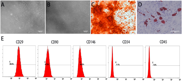
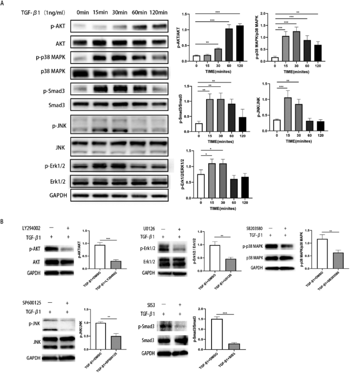
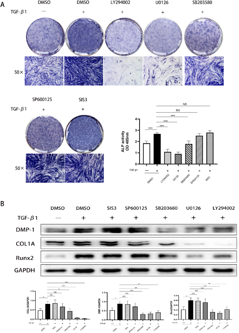
DPSCs were treated with 1 ng/mL TGF-β1 and Western blotting was conducted to examine the activation of protein kinase B (AKT), small mothers against decapentaplegic 3 (Smad3), p38 mitogen-activated protein kinase (p38 MAPK), c-Jun N-terminal kinase (JNK) and extracellular signal-regulated kinase 1/2 (Erk1/2). DPSCs were exposed to mineralization medium contained TGF-β1 with/without the specific signaling pathway inhibitors, and early odontogenic differentiation was evaluated by assessing the expression of alkaline phosphatase (ALP), collagen type 1 alpha 1 (COL1A), dentin matrix protein 1 (DMP-1) and runt-related transcription factor 2 (Runx2).
TGF-β1 stimulated AKT, Smad3, p38 MAPK, Erk1/2 and JNK phosphorylation in DPSCs within 120 min. TGF-β1 enhanced ALP activity and elevated levels of COL1A, DMP-1 and Runx2. LY294002, U0126 and SB203580 attenuated the effect of TGF-β1 on DPSCs, however, the SIS3 and SP600125 treated groups had no significant effect.
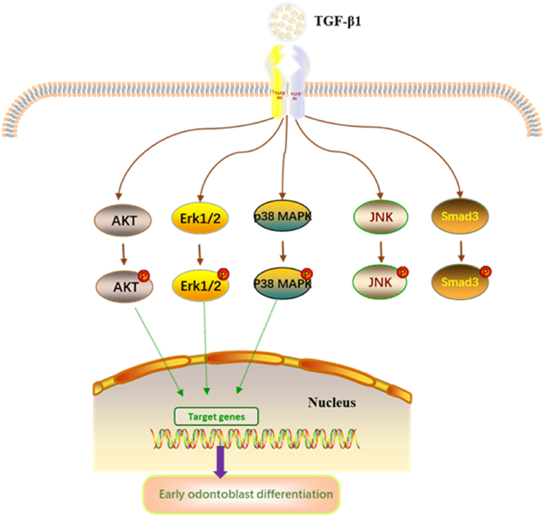
TGF-β1 promotes the early stage of odontoblastic differentiation in DPSCs by activating AKT, Erk1/2 and p38 MAPK signaling pathways, but not by Smad3 and JNK.
背景/目的:转化生长因子-β1(TGF-β1)在牙髓-牙本质复合体的再生和修复中起重要作用。然而,TGF-β1对成牙本质细胞分化的生物学功能仍不清楚,主要是因为分化过程受复杂信号通路控制。本研究旨在探讨TGF-β1调控牙髓干细胞(DPSCs)早期分化所涉及的信号通路及其功能作用。
用1 ng/mL TGF-β1处理DPSCs,进行蛋白质免疫印迹法检测蛋白激酶B(AKT)、小母细胞磷酸化蛋白3(Smad3)、p38丝裂原活化蛋白激酶(p38 MAPK)、c-Jun氨基末端激酶(JNK)和细胞外信号调节激酶1/2(Erk1/2)的激活情况。将DPSCs置于含或不含特定信号通路抑制剂的含TGF-β1的矿化培养基中,通过评估碱性磷酸酶(ALP)、Ⅰ型胶原蛋白α1(COL1A)、牙本质基质蛋白1(DMP-1)和 runt相关转录因子2(Runx2)的表达来评价早期成牙本质细胞分化。
TGF-β1在120分钟内刺激DPSCs中AKT、Smad3、p38 MAPK、Erk1/2和JNK磷酸化。TGF-β1增强了ALP活性,提高了COL1A、DMP-1和Runx2的水平。LY294002、U0126和SB203580减弱了TGF-β1对DPSCs的作用,然而,SIS3和SP600125处理组没有显著影响。
TGF-β1通过激活AKT、Erk1/2和p38 MAPK信号通路促进DPSCs成牙本质细胞分化的早期阶段,但不通过Smad3和JNK。