• 文献检索
  • 文档翻译
  • 深度研究
  • 学术资讯
  • Suppr Zotero 插件Zotero 插件
  • 邀请有礼
  • 套餐&价格
  • 历史记录
应用&插件
Suppr Zotero 插件Zotero 插件浏览器插件Mac 客户端Windows 客户端微信小程序
定价
高级版会员购买积分包购买API积分包
服务
文献检索文档翻译深度研究API 文档MCP 服务
关于我们
关于 Suppr公司介绍联系我们用户协议隐私条款
关注我们

Suppr 超能文献

核心技术专利:CN118964589B侵权必究
粤ICP备2023148730 号-1Suppr @ 2026

文献检索

告别复杂PubMed语法,用中文像聊天一样搜索,搜遍4000万医学文献。AI智能推荐,让科研检索更轻松。

立即免费搜索

文件翻译

保留排版,准确专业,支持PDF/Word/PPT等文件格式,支持 12+语言互译。

免费翻译文档

深度研究

AI帮你快速写综述,25分钟生成高质量综述,智能提取关键信息,辅助科研写作。

立即免费体验

使用人工智能对前列腺癌 PET/CT 骨骼肿瘤负荷进行自动定量:PET 指数。

Automated quantification of PET/CT skeletal tumor burden in prostate cancer using artificial intelligence: The PET index.

机构信息

Division of Nuclear Medicine, Department of Translational Medicine, Lund University, Malmö, Sweden.

Department of Surgery, Skåne University Hospital, Malmö, Sweden.

出版信息

Eur J Nucl Med Mol Imaging. 2023 Apr;50(5):1510-1520. doi: 10.1007/s00259-023-06108-4. Epub 2023 Jan 18.

DOI:10.1007/s00259-023-06108-4
PMID:36650356
原文链接:https://pmc.ncbi.nlm.nih.gov/articles/PMC10027829/
Abstract

PURPOSE

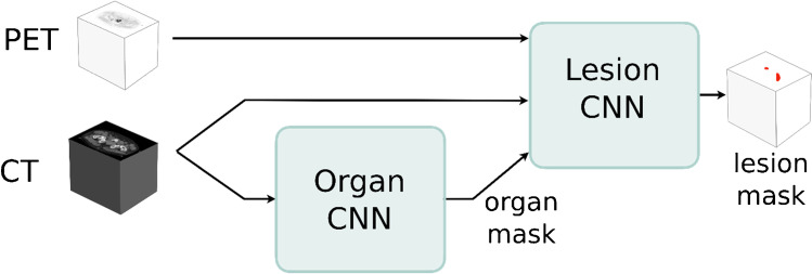
Consistent assessment of bone metastases is crucial for patient management and clinical trials in prostate cancer (PCa). We aimed to develop a fully automated convolutional neural network (CNN)-based model for calculating PET/CT skeletal tumor burden in patients with PCa.

METHODS

A total of 168 patients from three centers were divided into training, validation, and test groups. Manual annotations of skeletal lesions in [F]fluoride PET/CT scans were used to train a CNN. The AI model was evaluated in 26 patients and compared to segmentations by physicians and to a SUV 15 threshold. PET index representing the percentage of skeletal volume taken up by lesions was estimated.

RESULTS

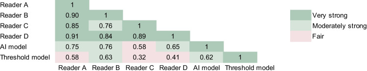
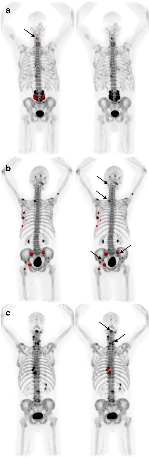
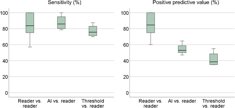
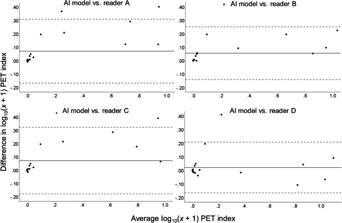
There was no case in which all readers agreed on prevalence of lesions that the AI model failed to detect. PET index by the AI model correlated moderately strong to physician PET index (mean r = 0.69). Threshold PET index correlated fairly with physician PET index (mean r = 0.49). The sensitivity for lesion detection was 65-76% for AI, 68-91% for physicians, and 44-51% for threshold depending on which physician was considered reference.

CONCLUSION

It was possible to develop an AI-based model for automated assessment of PET/CT skeletal tumor burden. The model's performance was superior to using a threshold and provides fully automated calculation of whole-body skeletal tumor burden. It could be further developed to apply to different radiotracers. Objective scan evaluation is a first step toward developing a PET/CT imaging biomarker for PCa skeletal metastases.

摘要

目的

对前列腺癌(PCa)患者的骨转移进行一致的评估对于患者管理和临床试验至关重要。我们旨在开发一种基于完全自动化卷积神经网络(CNN)的模型,用于计算 PCa 患者的 PET/CT 骨骼肿瘤负担。

方法

来自三个中心的 168 名患者被分为训练组、验证组和测试组。使用 [F]氟化物 PET/CT 扫描中的骨骼病变的手动注释来训练 CNN。该 AI 模型在 26 名患者中进行了评估,并与医生的分割以及 SUV15 阈值进行了比较。估计代表病变占据骨骼体积百分比的 PET 指数。

结果

没有一个病例是所有读者都同意 AI 模型未能检测到的病变的患病率。AI 模型的 PET 指数与医生的 PET 指数中度相关(平均 r=0.69)。阈值 PET 指数与医生的 PET 指数相当相关(平均 r=0.49)。对于 AI,对于医生,对于阈值,病变检测的敏感性分别为 65-76%、68-91%和 44-51%,具体取决于哪个医生被认为是参考。

结论

可以开发一种基于 AI 的模型,用于自动评估 PET/CT 骨骼肿瘤负担。该模型的性能优于使用阈值,并且可以提供全身骨骼肿瘤负担的全自动计算。它可以进一步开发以应用于不同的放射性示踪剂。客观扫描评估是开发用于 PCa 骨骼转移的 PET/CT 成像生物标志物的第一步。

https://cdn.ncbi.nlm.nih.gov/pmc/blobs/a35f/10027829/e50ed9b734d1/259_2023_6108_Fig5_HTML.jpg
https://cdn.ncbi.nlm.nih.gov/pmc/blobs/a35f/10027829/8d7354982cbe/259_2023_6108_Fig1_HTML.jpg
https://cdn.ncbi.nlm.nih.gov/pmc/blobs/a35f/10027829/f0f94606a216/259_2023_6108_Fig2_HTML.jpg
https://cdn.ncbi.nlm.nih.gov/pmc/blobs/a35f/10027829/1063fc117281/259_2023_6108_Fig3_HTML.jpg
https://cdn.ncbi.nlm.nih.gov/pmc/blobs/a35f/10027829/23db88ed4da0/259_2023_6108_Fig4_HTML.jpg
https://cdn.ncbi.nlm.nih.gov/pmc/blobs/a35f/10027829/e50ed9b734d1/259_2023_6108_Fig5_HTML.jpg

相似文献

1
Automated quantification of PET/CT skeletal tumor burden in prostate cancer using artificial intelligence: The PET index.使用人工智能对前列腺癌 PET/CT 骨骼肿瘤负荷进行自动定量:PET 指数。
Eur J Nucl Med Mol Imaging. 2023 Apr;50(5):1510-1520. doi: 10.1007/s00259-023-06108-4. Epub 2023 Jan 18.
2
Deep learning for segmentation of 49 selected bones in CT scans: First step in automated PET/CT-based 3D quantification of skeletal metastases.深度学习在 CT 扫描中对 49 块选定骨骼的分割:基于 PET/CT 的骨骼转移全自动 3D 定量的第一步。
Eur J Radiol. 2019 Apr;113:89-95. doi: 10.1016/j.ejrad.2019.01.028. Epub 2019 Feb 1.
3
More advantages in detecting bone and soft tissue metastases from prostate cancer using F-PSMA PET/CT.使用F-PSMA PET/CT检测前列腺癌骨和软组织转移方面有更多优势。
Hell J Nucl Med. 2019 Jan-Apr;22(1):6-9. doi: 10.1967/s002449910952. Epub 2019 Mar 7.
4
Freely available artificial intelligence for pelvic lymph node metastases in PSMA PET-CT that performs on par with nuclear medicine physicians.可自由获取的用于 PSMA PET-CT 中盆腔淋巴结转移的人工智能,其表现与核医学医师相当。
Eur J Nucl Med Mol Imaging. 2022 Aug;49(10):3412-3418. doi: 10.1007/s00259-022-05806-9. Epub 2022 Apr 27.
5
Artificial intelligence-based detection of lymph node metastases by PET/CT predicts prostate cancer-specific survival.基于人工智能的 PET/CT 检测淋巴结转移可预测前列腺癌特异性生存。
Clin Physiol Funct Imaging. 2021 Jan;41(1):62-67. doi: 10.1111/cpf.12666. Epub 2020 Oct 18.
6
Deep learning-based quantification of PET/CT prostate gland uptake: association with overall survival.基于深度学习的PET/CT前列腺摄取定量分析:与总生存期的关联
Clin Physiol Funct Imaging. 2020 Mar;40(2):106-113. doi: 10.1111/cpf.12611. Epub 2019 Dec 20.
7
Freely Available, Fully Automated AI-Based Analysis of Primary Tumour and Metastases of Prostate Cancer in Whole-Body [F]-PSMA-1007 PET-CT.基于人工智能的全身[F]-PSMA-1007 PET-CT前列腺癌原发肿瘤和转移灶免费且全自动分析
Diagnostics (Basel). 2022 Aug 30;12(9):2101. doi: 10.3390/diagnostics12092101.
8
Automatic segmentation of prostate cancer metastases in PSMA PET/CT images using deep neural networks with weighted batch-wise dice loss.使用带有加权批次骰子损失的深度神经网络对PSMA PET/CT图像中的前列腺癌转移灶进行自动分割。
Comput Biol Med. 2023 May;158:106882. doi: 10.1016/j.compbiomed.2023.106882. Epub 2023 Apr 4.
9
Fully automatic prognostic biomarker extraction from metastatic prostate lesion segmentations in whole-body [Ga]Ga-PSMA-11 PET/CT images.从全身[Ga]Ga-PSMA-11 PET/CT 图像中转移性前列腺病变分割中全自动提取预后生物标志物。
Eur J Nucl Med Mol Imaging. 2022 Dec;50(1):67-79. doi: 10.1007/s00259-022-05927-1. Epub 2022 Aug 17.
10
Artificial intelligence-based versus manual assessment of prostate cancer in the prostate gland: a method comparison study.基于人工智能与人工评估前列腺中前列腺癌的比较研究
Clin Physiol Funct Imaging. 2019 Nov;39(6):399-406. doi: 10.1111/cpf.12592. Epub 2019 Sep 8.

引用本文的文献

1
Prediction of bone oligometastases in breast cancer using models based on deep learning radiomics of PET/CT imaging.使用基于PET/CT成像深度学习影像组学的模型预测乳腺癌骨寡转移。
Front Oncol. 2025 Aug 21;15:1621677. doi: 10.3389/fonc.2025.1621677. eCollection 2025.
2
Fully automated 3D multi-modal deep learning model for preoperative T-stage prediction of colorectal cancer using F-FDG PET/CT.用于使用F-FDG PET/CT进行结直肠癌术前T分期预测的全自动3D多模态深度学习模型。
Eur J Nucl Med Mol Imaging. 2025 Jul 28. doi: 10.1007/s00259-025-07450-5.
3
The Evolving Role of PSMA-PET/CT in Prostate Cancer Management: an Umbrella Review of Diagnostic Restaging, Therapeutic Redirection, and Survival Impact.

本文引用的文献

1
Freely Available, Fully Automated AI-Based Analysis of Primary Tumour and Metastases of Prostate Cancer in Whole-Body [F]-PSMA-1007 PET-CT.基于人工智能的全身[F]-PSMA-1007 PET-CT前列腺癌原发肿瘤和转移灶免费且全自动分析
Diagnostics (Basel). 2022 Aug 30;12(9):2101. doi: 10.3390/diagnostics12092101.
2
Fully automatic prognostic biomarker extraction from metastatic prostate lesion segmentations in whole-body [Ga]Ga-PSMA-11 PET/CT images.从全身[Ga]Ga-PSMA-11 PET/CT 图像中转移性前列腺病变分割中全自动提取预后生物标志物。
Eur J Nucl Med Mol Imaging. 2022 Dec;50(1):67-79. doi: 10.1007/s00259-022-05927-1. Epub 2022 Aug 17.
3
PSMA-PET/CT在前列腺癌管理中不断演变的作用:关于诊断性再分期、治疗调整及生存影响的综合综述
Curr Oncol Rep. 2025 May 14. doi: 10.1007/s11912-025-01682-2.
4
Oncologic Applications of Artificial Intelligence and Deep Learning Methods in CT Spine Imaging-A Systematic Review.人工智能和深度学习方法在CT脊柱成像中的肿瘤学应用——一项系统综述
Cancers (Basel). 2024 Aug 28;16(17):2988. doi: 10.3390/cancers16172988.
Freely available artificial intelligence for pelvic lymph node metastases in PSMA PET-CT that performs on par with nuclear medicine physicians.
可自由获取的用于 PSMA PET-CT 中盆腔淋巴结转移的人工智能,其表现与核医学医师相当。
Eur J Nucl Med Mol Imaging. 2022 Aug;49(10):3412-3418. doi: 10.1007/s00259-022-05806-9. Epub 2022 Apr 27.
4
Analytical performance of aPROMISE: automated anatomic contextualization, detection, and quantification of [F]DCFPyL (PSMA) imaging for standardized reporting.aPROMISE 分析性能:用于标准化报告的 [F]DCFPyL(PSMA)成像的自动解剖语境化、检测和定量。
Eur J Nucl Med Mol Imaging. 2022 Feb;49(3):1041-1051. doi: 10.1007/s00259-021-05497-8. Epub 2021 Aug 31.
5
Whole-body uptake classification and prostate cancer staging in Ga-PSMA-11 PET/CT using dual-tracer learning.基于双示踪剂学习的 Ga-PSMA-11 PET/CT 全身摄取分类和前列腺癌分期。
Eur J Nucl Med Mol Imaging. 2022 Jan;49(2):517-526. doi: 10.1007/s00259-021-05473-2. Epub 2021 Jul 7.
6
E-PSMA: the EANM standardized reporting guidelines v1.0 for PSMA-PET.E-PSMA:EANM 标准化报告指南 v1.0 版用于 PSMA-PET。
Eur J Nucl Med Mol Imaging. 2021 May;48(5):1626-1638. doi: 10.1007/s00259-021-05245-y. Epub 2021 Feb 19.
7
RECOMIA-a cloud-based platform for artificial intelligence research in nuclear medicine and radiology.RECOMIA——一个用于核医学与放射学人工智能研究的基于云的平台。
EJNMMI Phys. 2020 Aug 4;7(1):51. doi: 10.1186/s40658-020-00316-9.
8
Consensus statements on PSMA PET/CT response assessment criteria in prostate cancer.前列腺癌 PSMA PET/CT 反应评估标准的共识声明。
Eur J Nucl Med Mol Imaging. 2021 Feb;48(2):469-476. doi: 10.1007/s00259-020-04934-4. Epub 2020 Jul 2.
9
Semiautomatically Quantified Tumor Volume Using Ga-PSMA-11 PET as a Biomarker for Survival in Patients with Advanced Prostate Cancer.使用 Ga-PSMA-11 PET 半自动量化肿瘤体积作为晚期前列腺癌患者生存的生物标志物。
J Nucl Med. 2020 Dec;61(12):1786-1792. doi: 10.2967/jnumed.120.242057. Epub 2020 Apr 24.
10
F-Sodium fluoride PET/CT predicts overall survival in patients with advanced genitourinary malignancies treated with cabozantinib and nivolumab with or without ipilimumab.F-氟化钠正电子发射断层扫描/计算机断层扫描(F-Sodium fluoride PET/CT)可预测接受卡博替尼和纳武单抗(无论是否联用伊匹木单抗)治疗的晚期泌尿生殖系统恶性肿瘤患者的总生存期。
Eur J Nucl Med Mol Imaging. 2020 Jan;47(1):178-184. doi: 10.1007/s00259-019-04483-5. Epub 2019 Sep 14.