Department of Neurosurgery, Charité-Universitätsmedizin Berlin, corporate member of Freie Universität Berlin, Humboldt-Universität Zu Berlin, Berlin Institute of Health, Berlin, Germany.
Department of Neurosurgery, Universitätsklinikum Hamburg-Eppendorf, Hamburg, Germany.
Acta Neurochir (Wien). 2023 Mar;165(3):771-777. doi: 10.1007/s00701-022-05470-w. Epub 2023 Jan 18.
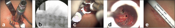
Thoracic disc herniations are uncommon and carry a high risk for neurological deterioration. Traditional surgical approaches include thoracotomy, costotransversectomy or posterior approaches with considerable morbidity. In this technical note with case series, we describe a minimally invasive tubular retractor-assisted retropleural approach for simple and less invasive microsurgical exploration of thoracic disc herniations from a lateral angle.
Surgical technique consisted of partial rib resection and retropleural dissection followed by the placement of a tubular retractor (METRx Tubes, Medtronic) for an anterior-lateral exposure of the disc and neuroforamen. Epidemiological, clinical and surgical patient data were acquired.
Between 2017 and 2020, six patients were surgically treated using the minimally invasive tubular retractor-assisted retropleural approach. Microsurgical exposure of the disc and neural structures was achieved from a lateral direction without requiring thoracotomy or lung deflation. Control imaging confirmed resection in all cases without relevant residuum. As postoperative complications, one dural injury and one postoperative pneumothorax occured. No neurologic deterioration or recurrence occurred during a median follow-up of 3 months.
The described tubular retractor-assisted retropleural exposure serves as a feasible minimally invasive microsurgical approach to the anterior-lateral thoracic spine.
胸椎间盘突出症并不常见,但存在神经功能恶化的高风险。传统的手术方法包括开胸术、肋横突切除术或后路手术,但这些方法的发病率较高。在本技术说明及病例系列中,我们描述了一种微创管状牵开器辅助经胸膜后入路,从侧方角对胸椎间盘突出症进行简单、微创的显微镜下探查。
手术技术包括部分肋骨切除和胸膜后解剖,然后放置管状牵开器(METRx 管,美敦力),从前外侧暴露椎间盘和神经孔。收集了流行病学、临床和手术患者的数据。
在 2017 年至 2020 年期间,使用微创管状牵开器辅助经胸膜后入路对 6 名患者进行了手术治疗。通过侧向方向进行了椎间盘和神经结构的显微镜下暴露,无需开胸或肺萎陷。术后影像学检查证实所有病例均达到了切除效果,无明显残留。术后并发症包括 1 例硬脑膜损伤和 1 例气胸。在 3 个月的中位随访期间,没有出现神经功能恶化或复发。
所描述的管状牵开器辅助经胸膜后入路是一种可行的微创显微镜下治疗前外侧胸椎的方法。