Department of Pulmonary Medicine, FirstHealth of the Carolinas & Pinehurst Medical Clinic, Pinehurst, North Carolina, USA.
Division of Thoracic Surgery, Department of Surgery, Mayo Clinic, Rochester, Minnesota, USA.
Respiration. 2023;102(3):182-193. doi: 10.1159/000528820. Epub 2023 Jan 18.
Image-guided percutaneous thermal ablation is an established treatment option for early-stage lung cancer in medically inoperable patients but carries a high risk of pleura-related complications, particularly pneumothorax.
This study aimed to determine if image-guided transbronchial microwave ablation (tMWA) is a feasible approach to treat peripheral stage 1 lung cancer.
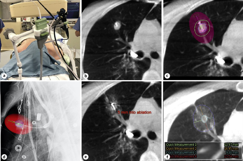
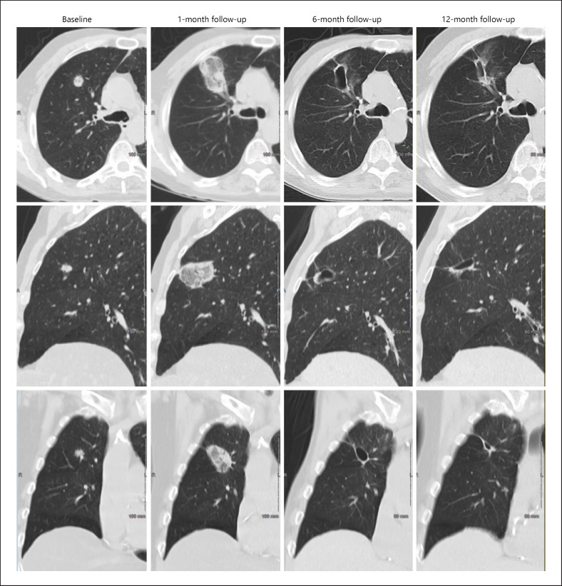
A prospective, single-arm, multicenter study sought to enroll 40 adults who were medically inoperable or declined surgery for peripheral stage 1 lung tumors (≤20 mm). Ablation was performed using navigational bronchoscopy and a flexible MWA probe, guided by cone-beam CT with augmented fluoroscopy. Follow-up at 1, 6, and 12 months included CT imaging of the ablation zone and possible tumor recurrence, adverse events (AEs), pulmonary function, and quality of life.
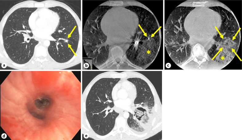
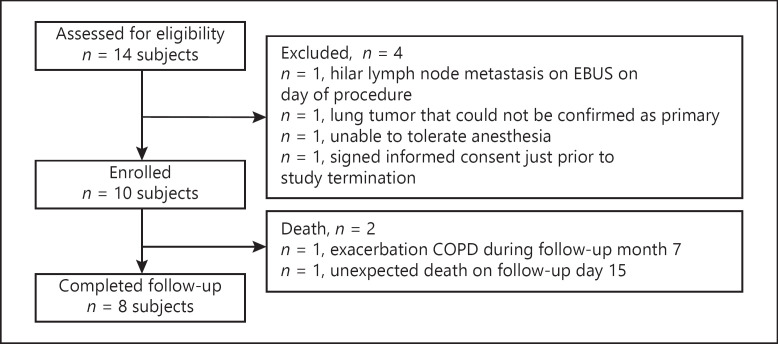
Across 2 sites, 11 tumors (10 NSCLC, 1 carcinoid) were treated in 10 enrolled patients. Median tumor diameter was 13 × 14 mm (7-19 mm) and median minimum ablative margin was 11 mm (5-19 mm). Technical success and technique efficacy were achieved in all patients. No tumor recurrence was seen during 12-month follow-up. No pneumothorax, pleural effusion, or bronchopleural fistula were noted. Minor AEs included scant hemoptysis, pain, cough, and dyspnea. Two serious AEs occurred ≤30 days of ablation and included a COPD exacerbation (day 9) and a death of unknown cause (day 15). The death led the sponsor to halt enrollment. Pulmonary function and quality-of-life indices remained stable.
Image-guided tMWA is a technically feasible approach for peripheral early-stage lung cancer but warrants further evaluation of safety and efficacy in larger cohorts.
影像引导经皮热消融是一种治疗不能手术的早期肺癌患者的成熟治疗方法,但存在较高的胸膜相关并发症风险,尤其是气胸。
本研究旨在确定影像引导经支气管微波消融(tMWA)是否是治疗外周 1 期肺癌的可行方法。
一项前瞻性、单臂、多中心研究旨在招募 40 名因医学原因无法手术或拒绝手术的外周 1 期肺肿瘤(≤20mm)的成年人。消融采用导航支气管镜和柔性 MWA 探头进行,在锥形束 CT 增强透视引导下进行。1、6 和 12 个月的随访包括消融区域的 CT 成像和可能的肿瘤复发、不良事件(AE)、肺功能和生活质量。
在 2 个地点,10 名入组患者的 11 个肿瘤(10 个 NSCLC,1 个类癌)接受了治疗。肿瘤直径中位数为 13×14mm(7-19mm),最小消融边界中位数为 11mm(5-19mm)。所有患者均达到了技术成功和技术疗效。12 个月随访期间未发现肿瘤复发。未发生气胸、胸腔积液或支气管胸膜瘘。轻微 AE 包括少量咯血、疼痛、咳嗽和呼吸困难。2 例严重 AE 在消融后 30 天内发生,包括 1 例 COPD 恶化(第 9 天)和 1 例死因不明的死亡(第 15 天)。该死亡导致赞助商停止入组。肺功能和生活质量指数保持稳定。
影像引导 tMWA 是治疗外周早期肺癌的一种技术可行的方法,但需要进一步评估更大队列的安全性和疗效。