Yoon Eun Jin, Lee Jun-Young, Kwak Seyul, Kim Yu Kyeong
Memory Network Medical Research Center, Seoul National University, Seoul, South Korea.
Department of Nuclear Medicine, SMG-SNU Boramae Medical Center, Seoul, South Korea.
Front Aging Neurosci. 2023 Jan 4;14:1051621. doi: 10.3389/fnagi.2022.1051621. eCollection 2022.
Mild behavioral impairment (MBI) is a neurobehavioral syndrome characterized by later life emergence of sustained neuropsychiatric symptoms, as an at-risk state for dementia. However, the associations between MBI and a risk of progression to Alzheimer's disease (AD) and its neuroanatomical correlates in mild cognitive impairment (MCI) are still unclear.
A total 1,184 older adults with amnestic MCI was followed for a mean of 3.1 ± 2.0 years. MBI was approximated using a transformation algorithm for the Neuropsychiatric Inventory at baseline. A two-step cluster analysis was used to identify subgroups of individuals with amnestic MCI based on profiles of 5 MBI domain symptoms (decreased motivation, affective dysregulation, impulse dyscontrol, social inappropriateness, abnormal perception/thought content). A Cox regression analysis was applied to investigate differences in the risk of progression to AD between subgroups. A subset of participants ( = 202) underwent 3D T1-weighted MRI scans at baseline and cortical thickness was compared between the subgroups of amnestic MCI patients.
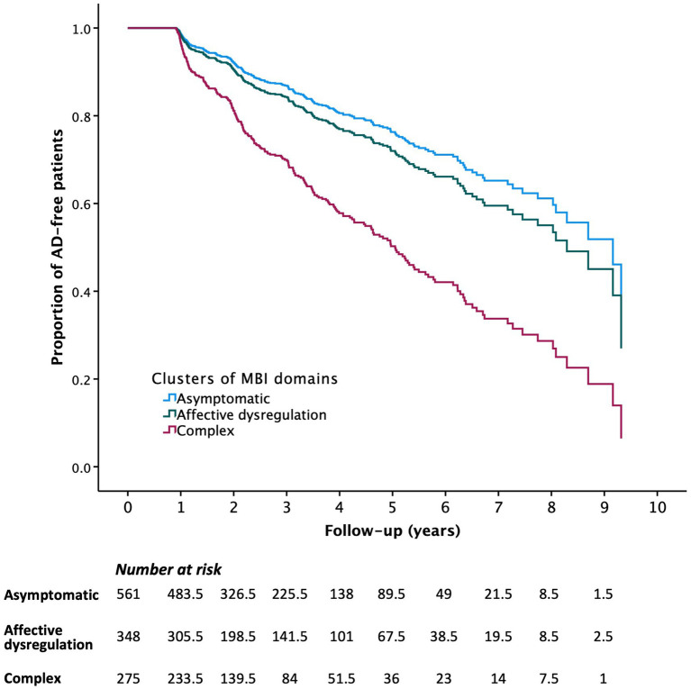
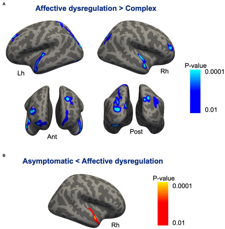
The cluster analysis classified the patients into 3 groups: (1) patients without any MBI domain symptoms (47.4%, asymptomatic group); (2) those with only affective dysregulation (29.4%, affective dysregulation group); (3) those with multiple MBI domain symptoms, particularly affective dysregulation, decreased motivation and impulse dyscontrol (23.2%, complex group). Compared to the asymptomatic group, the complex group was associated with a higher risk of progression to AD (hazard ratio = 2.541 [1.904-3.392], < 0.001), but the affective dysregulation group was not (1.214 [0.883-1.670], = 0.232). In cortical thickness analysis, the complex group revealed cortical thinning bilaterally in the inferior parietal, lateral occipital, lateral superior temporal, and frontopolar regions compared with the affective dysregulation group.
The multiple co-occuring MBI domains in individuals with amnestic MCI are associated with a higher risk of progression to AD and cortical thinning in temporal, parietal and frontal areas. These results suggest that evaluation of MBI could be useful for risk stratification for AD and appropriate intervention in MCI individuals.
轻度行为损害(MBI)是一种神经行为综合征,其特征是在晚年出现持续的神经精神症状,是痴呆症的风险状态。然而,MBI与进展为阿尔茨海默病(AD)的风险及其在轻度认知障碍(MCI)中的神经解剖学相关性仍不清楚。
对1184名遗忘型MCI的老年人进行了平均3.1±2.0年的随访。在基线时使用神经精神科问卷的转换算法估算MBI。采用两步聚类分析,根据5种MBI领域症状(动机减退、情感失调、冲动控制障碍、社交不当、异常感知/思维内容)的特征,识别遗忘型MCI个体的亚组。应用Cox回归分析来研究亚组之间进展为AD的风险差异。一部分参与者(n = 202)在基线时接受了3D T1加权MRI扫描,并比较了遗忘型MCI患者亚组之间的皮质厚度。
聚类分析将患者分为3组:(1)无任何MBI领域症状的患者(47.4%,无症状组);(2)仅患有情感失调的患者(29.4%,情感失调组);(3)患有多种MBI领域症状,尤其是情感失调、动机减退和冲动控制障碍的患者(23.2%,复杂组)。与无症状组相比,复杂组进展为AD的风险更高(风险比 = 2.541 [1.904 - 3.392],P < 0.001),但情感失调组并非如此(1.214 [0.883 - 1.670],P = 0.232)。在皮质厚度分析中,与情感失调组相比,复杂组在双侧下顶叶、枕外侧、颞上外侧和额极区域显示皮质变薄。
遗忘型MCI个体中多种同时出现的MBI领域与进展为AD的风险增加以及颞叶、顶叶和额叶区域的皮质变薄有关。这些结果表明,评估MBI可能有助于AD的风险分层以及对MCI个体进行适当干预。