Huaxi MR Research Center (HMRRC), Functional and Molecular Imaging Key Laboratory of Sichuan Province, Department of Radiology, West China Hospital of Sichuan University, Chengdu, Sichuan, 610041, P. R. China.
Psychoradiology Research Unit of Chinese Academy of Medical Sciences (2018RU011), West China Hospital of Sichuan University, Chengdu, 610041, China.
Neuropsychopharmacology. 2020 Mar;45(4):703-712. doi: 10.1038/s41386-019-0563-9. Epub 2019 Nov 6.
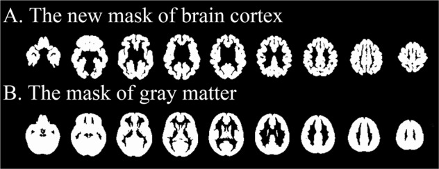
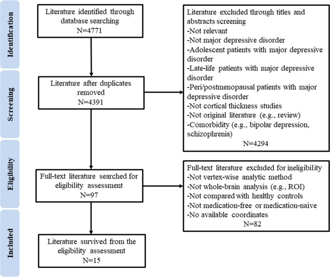
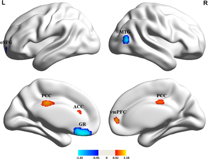
Alterations in cortical thickness have been identified in major depressive disorder (MDD), but findings have been variable and inconsistent. To date, no reliable tools have been available for the meta-analysis of surface-based morphometric (SBM) studies to effectively characterize what has been learned in previous studies, and drug treatments may have differentially impacted findings. We conducted a comprehensive meta-analysis of magnetic resonance imaging (MRI) studies that explored cortical thickness in medication-free patients with MDD, using a newly developed meta-analytic mask compatible with seed-based d mapping (SDM) meta-analytic software. We performed the meta-regression to explore the effects of demographics and clinical characteristics on variation in cortical thickness in MDD. Fifteen studies describing 529 patients and 586 healthy controls (HCs) were included. Medication-free patients with MDD, relative to HCs, showed a complex pattern of increased cortical thickness in some areas (posterior cingulate cortex, ventromedial prefrontal cortex, and anterior cingulate cortex) and decreased cortical thickness in others (gyrus rectus, orbital segment of the superior frontal gyrus, and middle temporal gyrus). Most findings in the whole sample analysis were confirmed in a meta-analysis of studies recruiting medication-naive patients. Using the new mask specifically developed for SBM studies, this SDM meta-analysis provides evidence for regional cortical thickness alterations in MDD, mainly involving increased cortical thickness in the default mode network and decreased cortical thickness in the orbitofrontal and temporal cortex.
大脑皮层厚度的改变已在重度抑郁症(MDD)中被识别,但研究结果存在差异且不一致。迄今为止,还没有可靠的工具可用于基于表面形态计量学(SBM)研究的荟萃分析,无法有效地描述以前研究的结果,且药物治疗可能对研究结果有不同的影响。我们使用新开发的与基于种子的弥散张量成像(SDM)荟萃分析软件兼容的荟萃分析蒙版,对探索 MDD 患者在未用药状态下大脑皮层厚度的磁共振成像(MRI)研究进行了全面的荟萃分析。我们进行了荟萃回归分析,以探索人口统计学和临床特征对 MDD 大脑皮层厚度变化的影响。有 15 项研究描述了 529 名患者和 586 名健康对照者(HCs),包括在内。与 HCs 相比,MDD 患者在未用药状态下显示出一些区域大脑皮层厚度增加(后扣带回皮质、腹内侧前额皮质和前扣带回皮质)和其他区域大脑皮层厚度减少(直回、额上回眶部和颞中回)的复杂模式。全样本分析中的大多数发现都在对招募未用药患者的研究进行的荟萃分析中得到了证实。使用专门为 SBM 研究开发的新蒙版,该 SDM 荟萃分析为 MDD 中存在区域性大脑皮层厚度改变提供了证据,主要涉及默认模式网络大脑皮层厚度增加和眶额和颞叶皮层大脑皮层厚度减少。