• 文献检索
  • 文档翻译
  • 深度研究
  • 学术资讯
  • Suppr Zotero 插件Zotero 插件
  • 邀请有礼
  • 套餐&价格
  • 历史记录
应用&插件
Suppr Zotero 插件Zotero 插件浏览器插件Mac 客户端Windows 客户端微信小程序
定价
高级版会员购买积分包购买API积分包
服务
文献检索文档翻译深度研究API 文档MCP 服务
关于我们
关于 Suppr公司介绍联系我们用户协议隐私条款
关注我们

Suppr 超能文献

核心技术专利:CN118964589B侵权必究
粤ICP备2023148730 号-1Suppr @ 2026

文献检索

告别复杂PubMed语法,用中文像聊天一样搜索,搜遍4000万医学文献。AI智能推荐,让科研检索更轻松。

立即免费搜索

文件翻译

保留排版,准确专业,支持PDF/Word/PPT等文件格式,支持 12+语言互译。

免费翻译文档

深度研究

AI帮你快速写综述,25分钟生成高质量综述,智能提取关键信息,辅助科研写作。

立即免费体验

使用 Fiji GliaMorph 工具包分析神经胶质细胞形态。

Glia Cell Morphology Analysis Using the Fiji GliaMorph Toolkit.

机构信息

Institute of Ophthalmology, University College London, Greater London, UK.

出版信息

Curr Protoc. 2023 Jan;3(1):e654. doi: 10.1002/cpz1.654.

DOI:10.1002/cpz1.654
PMID:36688682
原文链接:https://pmc.ncbi.nlm.nih.gov/articles/PMC10108223/
Abstract

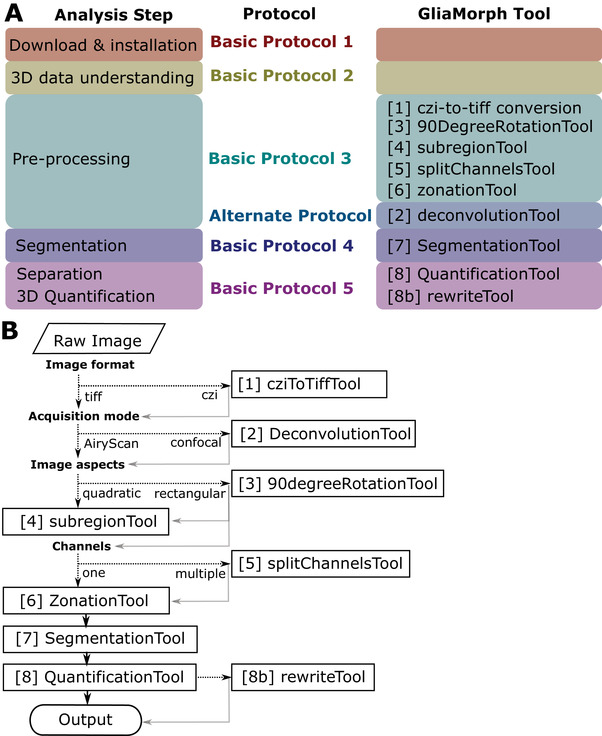
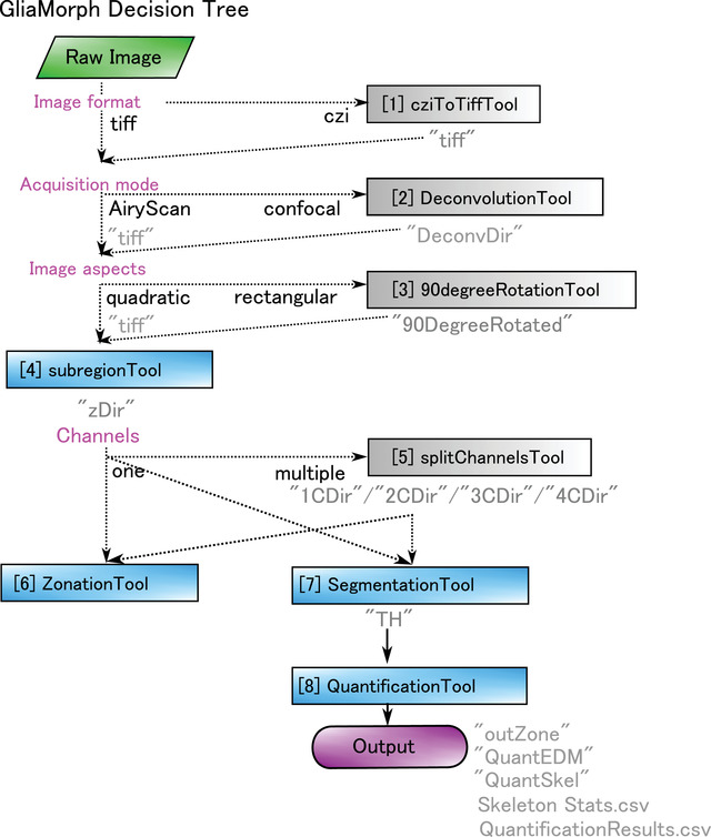
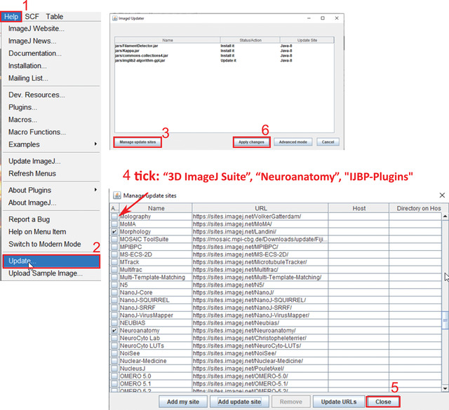
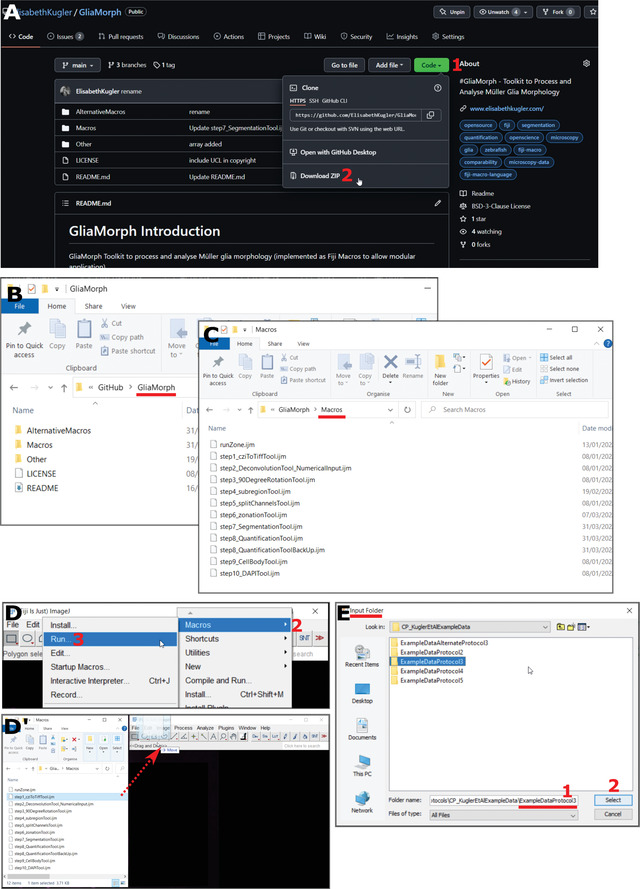
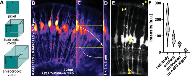
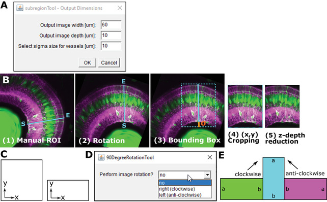
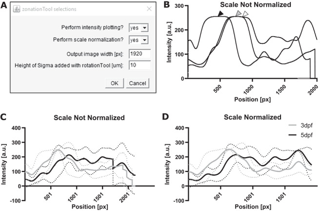
Glial cells are the support cells of the nervous system. Glial cells typically have elaborate morphologies that facilitate close contacts with neighboring neurons, synapses, and the vasculature. In the retina, Müller glia (MG) are the principal glial cell type that supports neuronal function by providing a myriad of supportive functions via intricate cell morphologies and precise contacts. Thus, complex glial morphology is critical for glial function, but remains challenging to resolve at a sub-cellular level or reproducibly quantify in complex tissues. To address this issue, we developed GliaMorph as a Fiji-based macro toolkit that allows 3D glial cell morphology analysis in the developing and mature retina. As GliaMorph is implemented in a modular fashion, here we present guides to (a) setup of GliaMorph, (b) data understanding in 3D, including z-axis intensity decay and signal-to-noise ratio, (c) pre-processing data to enhance image quality, (d) performing and examining image segmentation, and (e) 3D quantification of MG features, including apicobasal texture analysis. To allow easier application, GliaMorph tools are supported with graphical user interfaces where appropriate, and example data are publicly available to facilitate adoption. Further, GliaMorph can be modified to meet users' morphological analysis needs for other glial or neuronal shapes. Finally, this article provides users with an in-depth understanding of data requirements and the workflow of GliaMorph. © 2023 The Authors. Current Protocols published by Wiley Periodicals LLC. Basic Protocol 1: Download and installation of GliaMorph components including example data Basic Protocol 2: Understanding data properties and quality 3D-essential for subsequent analysis and capturing data property issues early Basic Protocol 3: Pre-processing AiryScan microscopy data for analysis Alternate Protocol: Pre-processing confocal microscopy data for analysis Basic Protocol 4: Segmentation of glial cells Basic Protocol 5: 3D quantification of glial cell morphology.

摘要

神经胶质细胞是神经系统的支持细胞。神经胶质细胞通常具有精细的形态,便于与邻近神经元、突触和血管紧密接触。在视网膜中,Müller 胶质细胞 (MG) 是主要的胶质细胞类型,通过复杂的细胞形态和精确的接触提供多种支持功能来支持神经元功能。因此,复杂的胶质形态对于胶质功能至关重要,但在亚细胞水平上解析或在复杂组织中重现定量仍然具有挑战性。为了解决这个问题,我们开发了 GliaMorph,这是一个基于 Fiji 的宏工具包,允许在发育中和成熟的视网膜中进行 3D 胶质细胞形态分析。由于 GliaMorph 以模块化的方式实现,因此我们在此提供了以下指南:(a) GliaMorph 的设置,(b) 3D 数据的理解,包括 z 轴强度衰减和信噪比,(c) 预处理数据以增强图像质量,(d) 执行和检查图像分割,以及 (e) MG 特征的 3D 定量,包括顶底纹理分析。为了方便应用,GliaMorph 工具在适当的情况下都支持图形用户界面,并且提供了公共示例数据,以方便采用。此外,GliaMorph 可以进行修改,以满足用户对其他胶质或神经元形状的形态分析需求。最后,本文为用户提供了对 GliaMorph 的数据要求和工作流程的深入了解。© 2023 作者。Wiley Periodicals LLC 出版的《当代协议》。基本方案 1:下载和安装 GliaMorph 组件,包括示例数据 基本方案 2:理解数据属性和 3D 质量——这是后续分析的基础,并且可以及早捕获数据属性问题 基本方案 3:使用 AiryScan 显微镜数据进行分析的预处理 可选方案:使用共聚焦显微镜数据进行分析的预处理 基本方案 4:胶质细胞的分割 基本方案 5:胶质细胞形态的 3D 定量

https://cdn.ncbi.nlm.nih.gov/pmc/blobs/9669/10108223/9810b414bf92/CPZ1-3-0-g007.jpg
https://cdn.ncbi.nlm.nih.gov/pmc/blobs/9669/10108223/75d21956aaa1/CPZ1-3-0-g004.jpg
https://cdn.ncbi.nlm.nih.gov/pmc/blobs/9669/10108223/72c46f94d4a5/CPZ1-3-0-g002.jpg
https://cdn.ncbi.nlm.nih.gov/pmc/blobs/9669/10108223/b0ef4f8f62df/CPZ1-3-0-g003.jpg
https://cdn.ncbi.nlm.nih.gov/pmc/blobs/9669/10108223/e767bbdbf114/CPZ1-3-0-g008.jpg
https://cdn.ncbi.nlm.nih.gov/pmc/blobs/9669/10108223/4c79919c7c88/CPZ1-3-0-g009.jpg
https://cdn.ncbi.nlm.nih.gov/pmc/blobs/9669/10108223/fc38b1accd39/CPZ1-3-0-g001.jpg
https://cdn.ncbi.nlm.nih.gov/pmc/blobs/9669/10108223/5b522b6d39b4/CPZ1-3-0-g006.jpg
https://cdn.ncbi.nlm.nih.gov/pmc/blobs/9669/10108223/919bfef365a6/CPZ1-3-0-g005.jpg
https://cdn.ncbi.nlm.nih.gov/pmc/blobs/9669/10108223/c9cc287a81da/CPZ1-3-0-g011.jpg
https://cdn.ncbi.nlm.nih.gov/pmc/blobs/9669/10108223/c1c5c50cc918/CPZ1-3-0-g010.jpg
https://cdn.ncbi.nlm.nih.gov/pmc/blobs/9669/10108223/9810b414bf92/CPZ1-3-0-g007.jpg

相似文献

1
Glia Cell Morphology Analysis Using the Fiji GliaMorph Toolkit.使用 Fiji GliaMorph 工具包分析神经胶质细胞形态。
Curr Protoc. 2023 Jan;3(1):e654. doi: 10.1002/cpz1.654.
2
GliaMorph: a modular image analysis toolkit to quantify Müller glial cell morphology.Gliamorph:一种用于量化 Müller 胶质细胞形态的模块化图像分析工具包。
Development. 2023 Feb 15;150(3). doi: 10.1242/dev.201008. Epub 2023 Feb 2.
3
Excitatory neurotransmission activates compartmentalized calcium transients in Müller glia without affecting lateral process motility.兴奋性神经递质传递激活 Müller 胶质细胞的分隔钙瞬变,而不影响侧突运动。
Elife. 2021 Dec 16;10:e73202. doi: 10.7554/eLife.73202.
4
mA epitranscriptomic modification regulates neural progenitor-to-glial cell transition in the retina.mA 表观转录修饰调控视网膜神经祖细胞向神经胶质细胞的转变。
Elife. 2022 Dec 2;11:e79994. doi: 10.7554/eLife.79994.
5
The inhibitor of phagocytosis, O-phospho-L-serine, suppresses Müller glia proliferation and cone cell regeneration in the light-damaged zebrafish retina.吞噬作用抑制剂 O-磷酸丝氨酸可抑制光损伤斑马鱼视网膜中的 Muller 胶质细胞增殖和视锥细胞再生。
Exp Eye Res. 2010 Nov;91(5):601-12. doi: 10.1016/j.exer.2010.07.017. Epub 2010 Aug 7.
6
Glial modulation of synaptic transmission in the retina.视网膜中神经胶质细胞对突触传递的调节
Glia. 2004 Aug 15;47(3):268-274. doi: 10.1002/glia.20030.
7
A novel type of glial cell in the retina is stimulated by insulin-like growth factor 1 and may exacerbate damage to neurons and Müller glia.视网膜中的一种新型神经胶质细胞受到胰岛素样生长因子 1 的刺激,可能会加剧神经元和 Müller 胶质细胞的损伤。
Glia. 2010 Apr 15;58(6):633-49. doi: 10.1002/glia.20950.
8
The glia of the adult Drosophila nervous system.成年果蝇神经系统中的神经胶质细胞。
Glia. 2017 Apr;65(4):606-638. doi: 10.1002/glia.23115. Epub 2017 Jan 30.
9
Peripapillary glial cells in the chick retina: A special glial cell type expressing astrocyte, radial glia, neuron, and oligodendrocyte markers throughout development.鸡视网膜中的视乳头周围神经胶质细胞:一种特殊的神经胶质细胞类型,在整个发育过程中表达星形胶质细胞、放射状胶质细胞、神经元和少突胶质细胞标志物。
Glia. 2004 May;46(4):346-55. doi: 10.1002/glia.10351.
10
Inhibition of Müller glial cell division blocks regeneration of the light-damaged zebrafish retina.抑制米勒胶质细胞分裂会阻碍光损伤斑马鱼视网膜的再生。
Dev Neurobiol. 2008 Feb 15;68(3):392-408. doi: 10.1002/dneu.20596.

引用本文的文献

1
Bone-derived PDGF-BB enhances hippocampal non-specific transcytosis through microglia-endothelial crosstalk in HFD-induced metabolic syndrome.骨衍生的 PDGF-BB 通过高脂肪饮食诱导的代谢综合征中的小胶质细胞-血管内皮细胞串扰增强海马非特异性转胞吞作用。
J Neuroinflammation. 2024 Apr 29;21(1):111. doi: 10.1186/s12974-024-03097-5.
2
GliaMorph: a modular image analysis toolkit to quantify Müller glial cell morphology.Gliamorph:一种用于量化 Müller 胶质细胞形态的模块化图像分析工具包。
Development. 2023 Feb 15;150(3). doi: 10.1242/dev.201008. Epub 2023 Feb 2.

本文引用的文献

1
GliaMorph: a modular image analysis toolkit to quantify Müller glial cell morphology.Gliamorph:一种用于量化 Müller 胶质细胞形态的模块化图像分析工具包。
Development. 2023 Feb 15;150(3). doi: 10.1242/dev.201008. Epub 2023 Feb 2.
2
Reactive astrocyte nomenclature, definitions, and future directions.反应性星形胶质细胞命名、定义和未来方向。
Nat Neurosci. 2021 Mar;24(3):312-325. doi: 10.1038/s41593-020-00783-4. Epub 2021 Feb 15.
3
Hypothesis-driven quantitative fluorescence microscopy - the importance of reverse-thinking in experimental design.
假说驱动的定量荧光显微镜 - 反向思维在实验设计中的重要性。
J Cell Sci. 2020 Nov 5;133(21):jcs250027. doi: 10.1242/jcs.250027.
4
Comparison of Multiscale Imaging Methods for Brain Research.多尺度成像方法在脑研究中的比较。
Cells. 2020 Jun 1;9(6):1377. doi: 10.3390/cells9061377.
5
Leveraging Zebrafish to Study Retinal Degenerations.利用斑马鱼研究视网膜变性。
Front Cell Dev Biol. 2018 Sep 19;6:110. doi: 10.3389/fcell.2018.00110. eCollection 2018.
6
A journey into the retina: Müller glia commanding survival and death.视网膜之旅:Müller 胶质细胞指挥着生存与死亡。
Eur J Neurosci. 2018 Jun;47(12):1429-1443. doi: 10.1111/ejn.13965. Epub 2018 Jun 8.
7
Glial control of neurogenesis.神经发生的神经胶质控制。
Curr Opin Neurobiol. 2017 Dec;47:188-195. doi: 10.1016/j.conb.2017.10.025. Epub 2017 Nov 13.
8
Cell Biology of Astrocyte-Synapse Interactions.星形胶质细胞-突触相互作用的细胞生物学
Neuron. 2017 Nov 1;96(3):697-708. doi: 10.1016/j.neuron.2017.09.056.
9
Mechanisms of Müller glial cell morphogenesis.Müller 胶质细胞形态发生的机制。
Curr Opin Neurobiol. 2017 Dec;47:31-37. doi: 10.1016/j.conb.2017.08.005. Epub 2017 Aug 29.
10
New astroglial injury-defined biomarkers for neurotrauma assessment.用于神经创伤评估的新的星形胶质细胞损伤定义生物标志物。
J Cereb Blood Flow Metab. 2017 Oct;37(10):3278-3299. doi: 10.1177/0271678X17724681. Epub 2017 Aug 17.