Institute of Biochemistry I, Jena University Hospital-Friedrich Schiller University Jena, Nonnenplan 2-4, 07743 Jena, Germany.
Core Facility Imaging, Leibniz Institute on Aging-Fritz Lipmann Institute (FLI), Beutenbergstraße 11, 07745 Jena, Germany.
Cells. 2020 Jun 1;9(6):1377. doi: 10.3390/cells9061377.
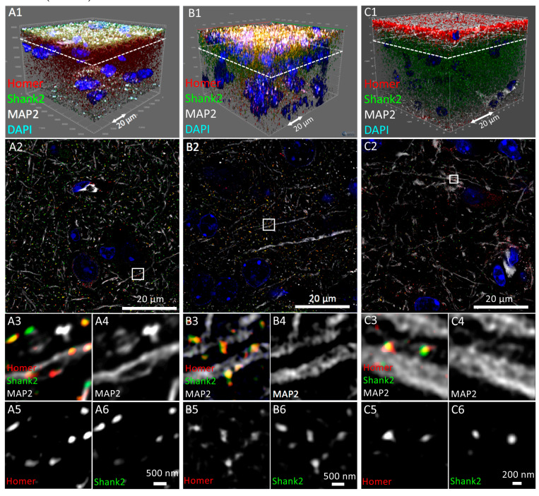
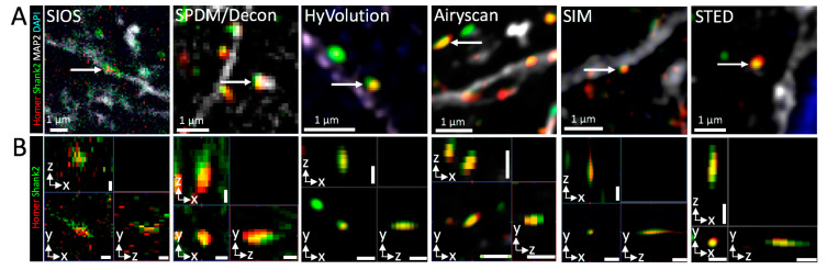
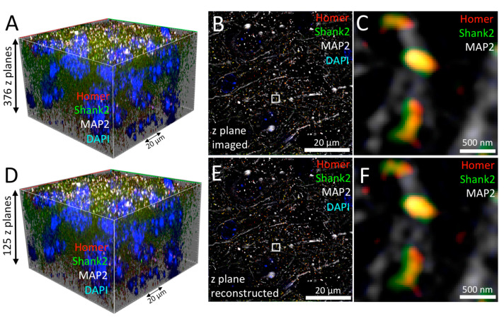
A major challenge in neuroscience is how to study structural alterations in the brain. Even small changes in synaptic composition could have severe outcomes for body functions. Many neuropathological diseases are attributable to disorganization of particular synaptic proteins. Yet, to detect and comprehensively describe and evaluate such often rather subtle deviations from the normal physiological status in a detailed and quantitative manner is very challenging. Here, we have compared side-by-side several commercially available light microscopes for their suitability in visualizing synaptic components in larger parts of the brain at low resolution, at extended resolution as well as at super-resolution. Microscopic technologies included stereo, widefield, deconvolution, confocal, and super-resolution set-ups. We also analyzed the impact of adaptive optics, a motorized objective correction collar and CUDA graphics card technology on imaging quality and acquisition speed. Our observations evaluate a basic set of techniques, which allow for multi-color brain imaging from centimeter to nanometer scales. The comparative multi-modal strategy we established can be used as a guide for researchers to select the most appropriate light microscopy method in addressing specific questions in brain research, and we also give insights into recent developments such as optical aberration corrections.
神经科学的一个主要挑战是如何研究大脑中的结构变化。即使是突触组成的微小变化也可能对身体功能产生严重影响。许多神经病理学疾病可归因于特定突触蛋白的紊乱。然而,以详细和定量的方式检测、全面描述和评估这种通常更为微妙的偏离正常生理状态的情况非常具有挑战性。在这里,我们并排比较了几种市售的光学显微镜,以评估它们在低分辨率、扩展分辨率和超分辨率下可视化大脑较大区域中突触成分的适用性。所使用的显微镜技术包括立体、宽场、反卷积、共聚焦和超分辨率设置。我们还分析了自适应光学、电动物镜校正环和 CUDA 显卡技术对成像质量和采集速度的影响。我们的观察结果评估了一套基本技术,这些技术允许从厘米到纳米尺度的多色大脑成像。我们建立的比较多模态策略可以作为研究人员在解决大脑研究中的具体问题时选择最合适的光学显微镜方法的指南,我们还深入探讨了像差校正等最新进展。