• 文献检索
  • 文档翻译
  • 深度研究
  • 学术资讯
  • Suppr Zotero 插件Zotero 插件
  • 邀请有礼
  • 套餐&价格
  • 历史记录
应用&插件
Suppr Zotero 插件Zotero 插件浏览器插件Mac 客户端Windows 客户端微信小程序
定价
高级版会员购买积分包购买API积分包
服务
文献检索文档翻译深度研究API 文档MCP 服务
关于我们
关于 Suppr公司介绍联系我们用户协议隐私条款
关注我们

Suppr 超能文献

核心技术专利:CN118964589B侵权必究
粤ICP备2023148730 号-1Suppr @ 2026

文献检索

告别复杂PubMed语法,用中文像聊天一样搜索,搜遍4000万医学文献。AI智能推荐,让科研检索更轻松。

立即免费搜索

文件翻译

保留排版,准确专业,支持PDF/Word/PPT等文件格式,支持 12+语言互译。

免费翻译文档

深度研究

AI帮你快速写综述,25分钟生成高质量综述,智能提取关键信息,辅助科研写作。

立即免费体验

对比增强乳腺摄影:重点关注常见的良性和恶性诊断。

Contrast enhanced mammography: focus on frequently encountered benign and malignant diagnoses.

机构信息

Department of Radiology, Cooper University Hospital, 1 Cooper Plaza, Camden, NJ, 08103, USA.

Present address: SimonMed Imaging, 6900 E Camelback Road, Suite 700, Scottsdale, AZ, 85251, USA.

出版信息

Cancer Imaging. 2023 Jan 23;23(1):10. doi: 10.1186/s40644-023-00526-1.

DOI:10.1186/s40644-023-00526-1
PMID:36691077
原文链接:https://pmc.ncbi.nlm.nih.gov/articles/PMC9872331/
Abstract

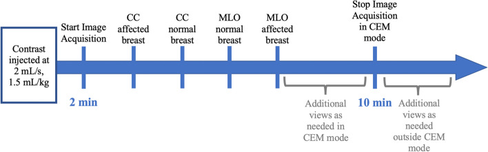
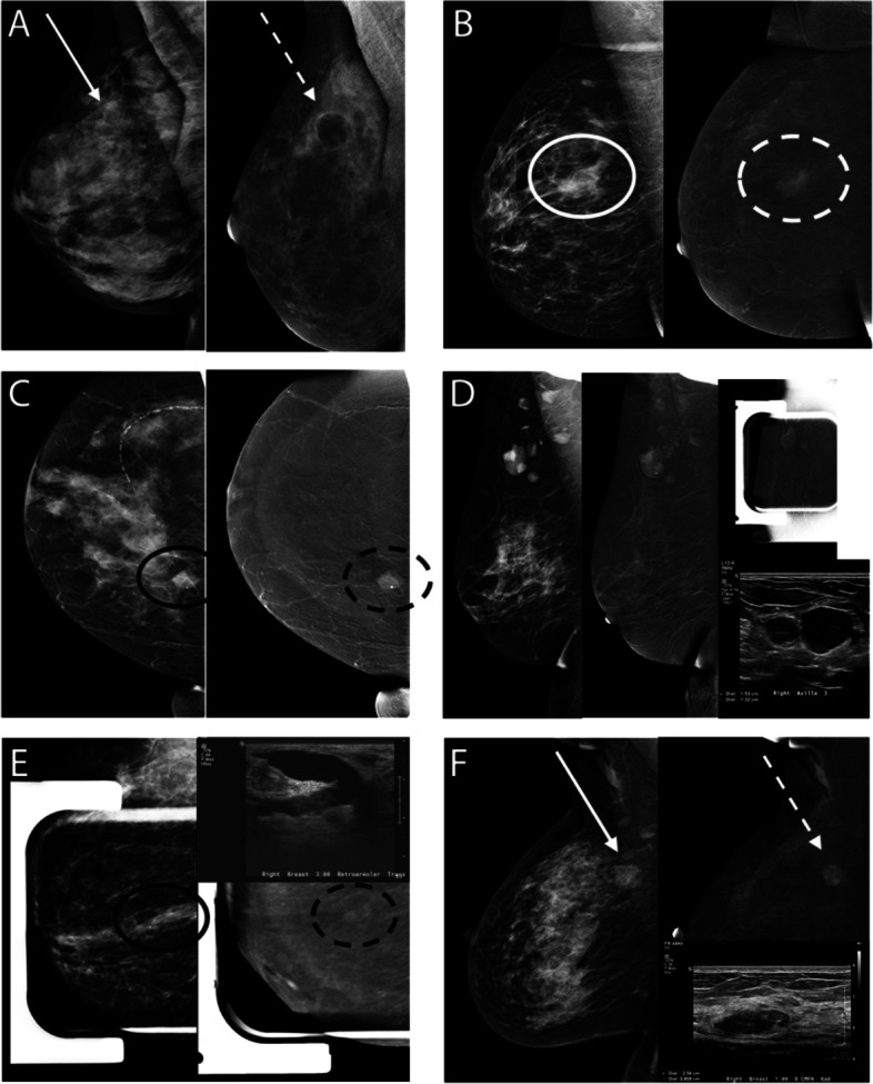
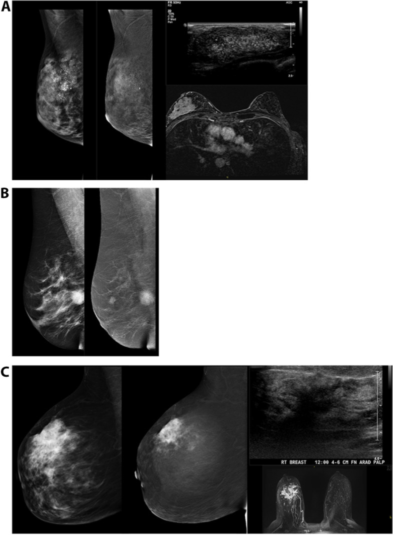
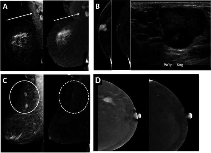
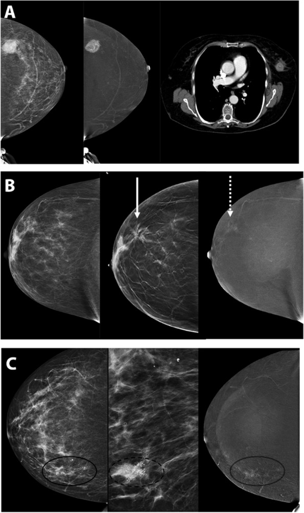
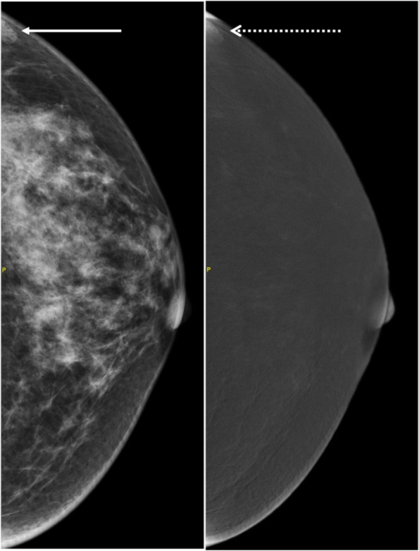
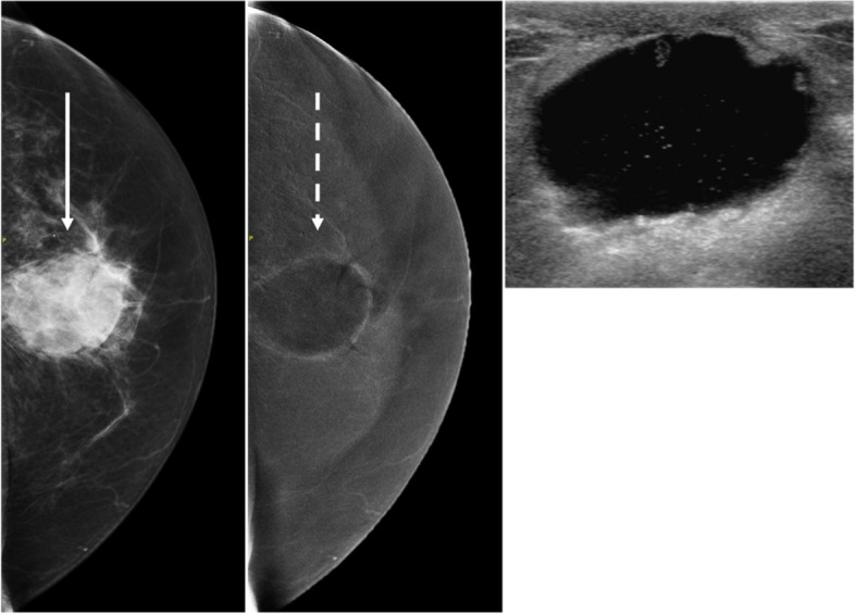
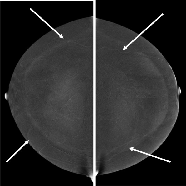
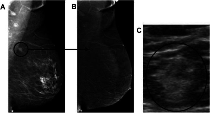
Contrast-enhanced mammography (CEM) is becoming a widely adopted modality in breast imaging over the past few decades and exponentially so over the last few years, with strong evidence of high diagnostic performance in cancer detection. Evidence is also growing indicating comparative performance of CEM to MRI in sensitivity with fewer false positive rates. As application of CEM ranges from potential use in screening dense breast populations to staging of known breast malignancy, increased familiarity with the modality and its implementation, and disease processes encountered becomes of great clinical significance. This review emphasizes expected normal findings on CEM followed by a focus on examples of the commonly encountered benign and malignant pathologies on CEM.

摘要

在过去几十年中,对比增强乳腺摄影(CEM)在乳腺成像中已被广泛采用,而且在过去几年中呈指数级增长,其在癌症检测方面具有出色的诊断性能。越来越多的证据表明,CEM 在敏感性方面与 MRI 的性能相当,假阳性率更低。由于 CEM 的应用范围从潜在用于筛查致密乳腺人群到已知乳腺恶性肿瘤的分期,因此对该模式及其应用以及所遇到的疾病过程的熟悉程度变得具有重要的临床意义。本综述强调了 CEM 上的预期正常发现,然后重点介绍了 CEM 上常见的良性和恶性病变的示例。

https://cdn.ncbi.nlm.nih.gov/pmc/blobs/112d/9872331/986b3d874cd0/40644_2023_526_Fig12_HTML.jpg
https://cdn.ncbi.nlm.nih.gov/pmc/blobs/112d/9872331/8662a7b52b4a/40644_2023_526_Fig1_HTML.jpg
https://cdn.ncbi.nlm.nih.gov/pmc/blobs/112d/9872331/4e286693aad0/40644_2023_526_Fig2_HTML.jpg
https://cdn.ncbi.nlm.nih.gov/pmc/blobs/112d/9872331/9d035186b03c/40644_2023_526_Fig3_HTML.jpg
https://cdn.ncbi.nlm.nih.gov/pmc/blobs/112d/9872331/c62673eb3933/40644_2023_526_Fig4_HTML.jpg
https://cdn.ncbi.nlm.nih.gov/pmc/blobs/112d/9872331/88cbf4467eaf/40644_2023_526_Fig5_HTML.jpg
https://cdn.ncbi.nlm.nih.gov/pmc/blobs/112d/9872331/fe210779ac14/40644_2023_526_Fig6_HTML.jpg
https://cdn.ncbi.nlm.nih.gov/pmc/blobs/112d/9872331/513f1f754c8d/40644_2023_526_Fig7_HTML.jpg
https://cdn.ncbi.nlm.nih.gov/pmc/blobs/112d/9872331/9c250a80a7ae/40644_2023_526_Fig8_HTML.jpg
https://cdn.ncbi.nlm.nih.gov/pmc/blobs/112d/9872331/74ee7eff31d7/40644_2023_526_Fig9_HTML.jpg
https://cdn.ncbi.nlm.nih.gov/pmc/blobs/112d/9872331/5a8609f668f8/40644_2023_526_Fig10_HTML.jpg
https://cdn.ncbi.nlm.nih.gov/pmc/blobs/112d/9872331/ec2d43ea44d0/40644_2023_526_Fig11_HTML.jpg
https://cdn.ncbi.nlm.nih.gov/pmc/blobs/112d/9872331/986b3d874cd0/40644_2023_526_Fig12_HTML.jpg

相似文献

1
Contrast enhanced mammography: focus on frequently encountered benign and malignant diagnoses.对比增强乳腺摄影:重点关注常见的良性和恶性诊断。
Cancer Imaging. 2023 Jan 23;23(1):10. doi: 10.1186/s40644-023-00526-1.
2
Comparison of Performance in Diagnosis and Characterization of Breast Lesions: Contrast-Enhanced Mammography Versus Breast Magnetic Resonance Imaging.对比增强乳腺摄影与乳腺磁共振成像在乳腺病变诊断和特征描述中的性能比较。
Clin Breast Cancer. 2024 Aug;24(6):481-493. doi: 10.1016/j.clbc.2024.04.007. Epub 2024 Apr 15.
3
Patient preferences regarding use of contrast-enhanced imaging for breast cancer screening.患者对乳腺癌筛查中使用对比增强成像的偏好。
Acad Radiol. 2022 Jan;29 Suppl 1:S229-S238. doi: 10.1016/j.acra.2021.03.003. Epub 2021 Apr 9.
4
Contrast-enhanced Mammography versus Contrast-enhanced Breast MRI: A Systematic Review and Meta-Analysis.对比增强乳腺 X 线摄影与对比增强乳腺 MRI:系统评价和荟萃分析。
Radiology. 2022 Oct;305(1):94-103. doi: 10.1148/radiol.212530. Epub 2022 Jun 7.
5
The PROCEM study protocol: Added value of preoperative contrast-enhanced mammography in staging of malignant breast lesions - a prospective randomized multicenter study.PROCEM 研究方案:术前对比增强乳腺摄影在恶性乳腺病变分期中的附加价值 - 一项前瞻性随机多中心研究。
BMC Cancer. 2021 Oct 18;21(1):1115. doi: 10.1186/s12885-021-08832-2.
6
Preoperative evaluation of breast cancer: Contrast-enhanced mammography versus contrast-enhanced magnetic resonance imaging: A systematic review and meta-analysis.术前乳腺癌评估:对比增强乳腺 X 线摄影与对比增强磁共振成像:系统评价和荟萃分析。
Breast Dis. 2022;41(1):303-315. doi: 10.3233/BD-210034.
7
Contrast-Enhanced Mammography Implementation, Performance, and Use for Supplemental Breast Cancer Screening.对比增强乳腺X线摄影术在补充性乳腺癌筛查中的实施、性能及应用
Radiol Clin North Am. 2021 Jan;59(1):113-128. doi: 10.1016/j.rcl.2020.08.006. Epub 2020 Oct 29.
8
Dynamic contrast-enhanced mammography and breast MRI in the diagnosis of breast cancer and detection of tumor size.动态对比增强乳腺摄影与乳腺 MRI 在乳腺癌诊断及肿瘤大小检测中的应用。
Turk J Med Sci. 2023 Dec 11;54(1):249-261. doi: 10.55730/1300-0144.5786. eCollection 2024.
9
Contrast-enhanced mammography in breast cancer screening.乳腺癌筛查中的对比增强乳腺摄影术。
Eur J Radiol. 2022 Nov;156:110513. doi: 10.1016/j.ejrad.2022.110513. Epub 2022 Sep 10.
10
Low-Dose, Contrast-Enhanced Mammography Compared to Contrast-Enhanced Breast MRI: A Feasibility Study.低剂量对比增强乳腺 X 光摄影与对比增强乳腺 MRI 对比:一项可行性研究。
J Magn Reson Imaging. 2020 Aug;52(2):589-595. doi: 10.1002/jmri.27079. Epub 2020 Feb 14.

引用本文的文献

1
Molecular and Biological Features of Invasive Lobular Carcinoma: Toward New Treatment Strategies.浸润性小叶癌的分子与生物学特征:迈向新的治疗策略
Ann Surg Oncol. 2025 Sep 12. doi: 10.1245/s10434-025-18259-9.
2
Contrast-Enhanced Mammography in Breast Lesion Assessment: Accuracy and Surgical Impact.对比增强乳腺钼靶在乳腺病变评估中的准确性及对手术的影响
Tomography. 2025 Aug 20;11(8):93. doi: 10.3390/tomography11080093.
3
Contrast-enhanced mammography-based interpretable machine learning model for the prediction of the molecular subtype breast cancers.

本文引用的文献

1
Contrast-Enhanced Mammography: A Scientific Review.对比增强乳腺X线摄影术:一项科学综述。
J Breast Imaging. 2020 Feb 4;2(1):7-15. doi: 10.1093/jbi/wbz074.
2
Radiation Doses and Risks in Breast Screening.乳腺筛查中的辐射剂量与风险
J Breast Imaging. 2020 Jun 3;2(3):188-200. doi: 10.1093/jbi/wbaa016.
3
Pearls and Pitfalls of Contrast-Enhanced Mammography.对比增强乳腺造影的要点与陷阱
基于对比增强乳腺X线摄影的可解释机器学习模型用于预测乳腺癌分子亚型
BMC Med Imaging. 2025 Jul 1;25(1):255. doi: 10.1186/s12880-025-01765-3.
4
Detection of a contralateral breast malignancy using contrast-enhanced mammography (CEM) overlooked by initial MRI scan.利用对比增强乳腺钼靶摄影(CEM)检测出初始MRI扫描遗漏的对侧乳腺恶性肿瘤。
Radiol Case Rep. 2025 Jun 11;20(9):4275-4285. doi: 10.1016/j.radcr.2025.05.046. eCollection 2025 Sep.
5
Diagnostic dilemma of lobular carcinoma: a mini-review of imaging modalities and the role of artificial intelligence and radiomics.小叶癌的诊断困境:影像学检查方法以及人工智能和放射组学作用的小型综述
Front Oncol. 2025 Mar 27;15:1515037. doi: 10.3389/fonc.2025.1515037. eCollection 2025.
6
Improving the diagnostic performance of contrast-enhanced mammography through lesion conspicuity and enhancement quantification.通过病变清晰度和增强量化提高对比增强乳腺摄影的诊断性能。
Eur Radiol. 2025 Apr 3. doi: 10.1007/s00330-025-11501-8.
7
Contrast-enhanced mammography improves patient access to functional breast imaging.对比增强乳腺钼靶摄影改善了患者获得功能性乳腺成像的机会。
J Med Imaging Radiat Oncol. 2025 Feb;69(1):46-61. doi: 10.1111/1754-9485.13789. Epub 2024 Oct 31.
8
Diagnostic Efficacy of Five Different Imaging Modalities in the Assessment of Women Recalled at Breast Screening-A Systematic Review and Meta-Analysis.五种不同成像方式在乳腺筛查召回女性评估中的诊断效能——一项系统评价与荟萃分析
Cancers (Basel). 2024 Oct 17;16(20):3505. doi: 10.3390/cancers16203505.
9
Role of contrast-enhanced mammogram as an adjunct to tomosynthesis in evaluation of circumscribed breast lesions.对比增强乳腺 X 线摄影在评估局限性乳腺病变中的作用。
Br J Radiol. 2024 Oct 1;97(1162):1696-1705. doi: 10.1093/bjr/tqae130.
10
Contrast-enhanced mammography BI-RADS: a case-based approach to radiology reporting.对比增强乳腺X线摄影BI-RADS:基于病例的放射学报告方法
Insights Imaging. 2024 Feb 8;15(1):37. doi: 10.1186/s13244-024-01612-z.
J Breast Imaging. 2019 Mar 13;1(1):64-72. doi: 10.1093/jbi/wby013.
4
Training Radiologists to Interpret Contrast-enhanced Mammography: Toward a Standardized Lexicon.培训放射科医生解读对比增强乳腺造影:迈向标准化词汇表
J Breast Imaging. 2021 Mar 20;3(2):176-189. doi: 10.1093/jbi/wbaa115.
5
Diagnostic Performance of Contrast-enhanced Mammography: Comparison With MRI and Mammography.对比增强乳腺X线摄影的诊断性能:与MRI和乳腺X线摄影的比较
J Breast Imaging. 2021 Aug 12;3(4):448-454. doi: 10.1093/jbi/wbab028.
6
Contrast-enhanced Mammography: A Guide to Setting Up a New Clinical Program.对比增强乳腺摄影:建立新临床项目指南
J Breast Imaging. 2021 May 21;3(3):369-376. doi: 10.1093/jbi/wbab027.
7
Clinical performance of contrast-enhanced spectral mammography in pre-surgical evaluation of breast malignant lesions in dense breasts: a single center study.对比增强光谱乳腺摄影术在致密型乳腺中乳腺恶性病变术前评估中的临床性能:一项单中心研究
Breast Cancer Res Treat. 2020 Dec;184(3):723-731. doi: 10.1007/s10549-020-05881-2. Epub 2020 Aug 28.
8
Artefacts in contrast enhanced digital mammography: how can they affect diagnostic image quality and confuse clinical diagnosis?对比增强数字乳腺摄影中的伪影:它们如何影响诊断图像质量并混淆临床诊断?
Insights Imaging. 2020 Feb 7;11(1):16. doi: 10.1186/s13244-019-0811-x.
9
Rapid Access to Contrast-Enhanced spectral mammogRaphy in women recalled from breast cancer screening: the RACER trial study design.从乳腺癌筛查中召回的女性的快速获取对比增强光谱 mammogRaphy:RACER 试验研究设计。
Trials. 2019 Dec 23;20(1):759. doi: 10.1186/s13063-019-3867-5.
10
Performance of Dual-Energy Contrast-enhanced Digital Mammography for Screening Women at Increased Risk of Breast Cancer.双能增强数字乳腺摄影筛查乳腺癌高危女性的性能。
Radiology. 2019 Oct;293(1):81-88. doi: 10.1148/radiol.2019182660. Epub 2019 Aug 27.