Department of Ophthalmology and Visual Sciences, Albert Einstein College of Medicine, Bronx, NY, 10461, USA.
Genetics, Albert Einstein College of Medicine, Bronx, NY, 10461, USA.
Epigenetics Chromatin. 2023 Jan 25;16(1):4. doi: 10.1186/s13072-023-00478-7.
Cellular differentiation is marked by temporally and spatially coordinated gene expression regulated at multiple levels. DNA methylation represents a universal mechanism to control chromatin organization and its accessibility. Cytosine methylation of CpG dinucleotides regulates binding of methylation-sensitive DNA-binding transcription factors within regulatory regions of transcription, including promoters and distal enhancers. Ocular lens differentiation represents an advantageous model system to examine these processes as lens comprises only two cell types, the proliferating lens epithelium and postmitotic lens fiber cells all originating from the epithelium.
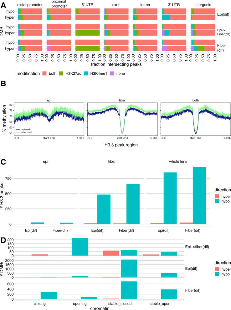
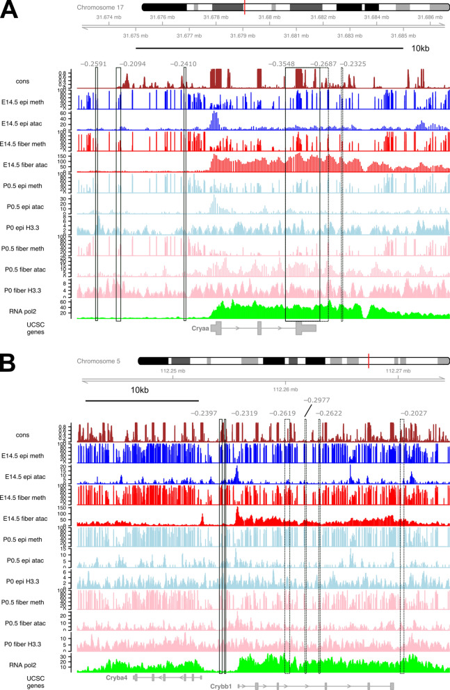
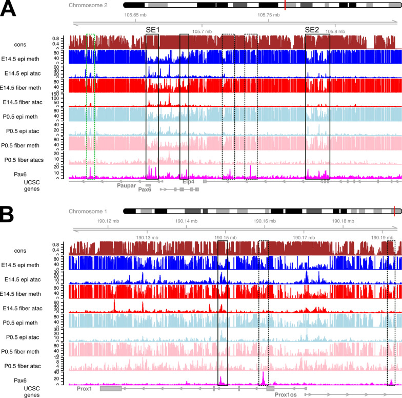
Using whole genome bisulfite sequencing (WGBS) and microdissected lenses, we investigated dynamics of DNA methylation and chromatin changes during mouse lens fiber and epithelium differentiation between embryos (E14.5) and newborns (P0.5). Histone H3.3 variant chromatin landscapes were also generated for both P0.5 lens epithelium and fibers by chromatin immunoprecipitation followed by next generation sequencing (ChIP-seq). Tissue-specific features of DNA methylation patterns are demonstrated via comparative studies with embryonic stem (ES) cells and neural progenitor cells (NPCs) at Nanog, Pou5f1, Sox2, Pax6 and Six3 loci. Comparisons with ATAC-seq and RNA-seq data demonstrate that reduced methylation is associated with increased expression of fiber cell abundant genes, including crystallins, intermediate filament (Bfsp1 and Bfsp2) and gap junction proteins (Gja3 and Gja8), marked by high levels of histone H3.3 within their transcribed regions. Interestingly, Pax6-binding sites exhibited predominantly DNA hypomethylation in lens chromatin. In vitro binding of Pax6 proteins showed Pax6's ability to interact with sites containing one or two methylated CpG dinucleotides.
Our study has generated the first data on methylation changes between two different stages of mammalian lens development and linked these data with chromatin accessibility maps, presence of histone H3.3 and gene expression. Reduced DNA methylation correlates with expression of important genes involved in lens morphogenesis and lens fiber cell differentiation.
细胞分化的特征是受多个水平调控的 temporally 和 spatially 协调的基因表达。DNA 甲基化是一种控制染色质组织及其可及性的通用机制。CpG 二核苷酸的胞嘧啶甲基化调节转录调控区域内甲基化敏感的 DNA 结合转录因子的结合,包括启动子和远端增强子。眼部晶状体分化是一个有利的模型系统,可用于研究这些过程,因为晶状体仅由两种细胞类型组成,即增殖的晶状体上皮细胞和有丝分裂后晶状体纤维细胞,它们均起源于上皮细胞。
我们使用全基因组亚硫酸氢盐测序 (WGBS) 和显微解剖的晶状体,研究了胚胎 (E14.5) 和新生 (P0.5) 之间小鼠晶状体纤维和上皮细胞分化过程中 DNA 甲基化和染色质变化的动态。我们还通过染色质免疫沉淀 followed by next generation sequencing (ChIP-seq) 为 P0.5 晶状体上皮细胞和纤维细胞生成了组蛋白 H3.3 变体染色质图谱。通过与胚胎干细胞 (ES 细胞) 和神经祖细胞 (NPCs) 进行比较研究,在 Nanog、Pou5f1、Sox2、Pax6 和 Six3 基因座上展示了 DNA 甲基化模式的组织特异性特征。与 ATAC-seq 和 RNA-seq 数据的比较表明,纤维细胞丰富基因的表达增加与甲基化减少相关,包括晶体蛋白、中间丝 (Bfsp1 和 Bfsp2) 和间隙连接蛋白 (Gja3 和 Gja8),其转录区域内存在高水平的组蛋白 H3.3。有趣的是,Pax6 结合位点在晶状体染色质中表现出主要的 DNA 低甲基化。体外 Pax6 蛋白结合实验表明 Pax6 能够与含有一个或两个甲基化 CpG 二核苷酸的位点相互作用。
我们的研究生成了哺乳动物晶状体发育两个不同阶段之间甲基化变化的首批数据,并将这些数据与染色质可及性图谱、组蛋白 H3.3 的存在和基因表达联系起来。DNA 去甲基化与参与晶状体形态发生和晶状体纤维细胞分化的重要基因的表达相关。