Forensic and Scientific Services, Health Support Queensland, Gold Coast University Hospital, Southport, QLD, Australia.
Griffith University School of Medicine, Southport, QLD, Australia.
Forensic Sci Med Pathol. 2023 Dec;19(4):479-483. doi: 10.1007/s12024-023-00579-5. Epub 2023 Jan 27.
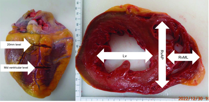
Cardiac ventricular dimensions measured at postmortem examination are used to assess whether there is hypertrophy of the heart chambers. However, there is no clear consensus on where these measurements should be taken. Some have proposed this should be measured at the mid-ventricular level, but others advocate it should be measured at a set distance (e.g. 20 mm) from the base of the heart. Twenty consecutive adult hearts were examined and showed the ventricular dimensions were significantly higher (mean: 5-15 mm, p < 0.01) when measured at a level 20 mm from the base of the heart compared to the mid-ventricular level. Of clinical significance is that in slightly less than half the cases, normal ventricular dimensions at mid ventricle level fell within the criteria considered pathological (> 40 mm) when measured at 20 mm from the base of the heart. In terms of actual ventricular dimensions, only the left ventricle diameter measured at 20 mm from the base of the heart correlated significantly (albeit moderately) with heart weight, suggesting it can be a predictor for cardiac hypertrophy.
心脏心室在尸检时测量的尺寸用于评估心脏腔室是否有肥大。然而,对于应该在哪里进行这些测量,目前还没有明确的共识。有人建议应该在心室中部进行测量,但也有人主张应该在距心脏底部一定距离(例如 20 毫米)处进行测量。对连续 20 例成人心脏进行检查,结果显示,与心室中部水平相比,距心脏底部 20 毫米处测量的心室尺寸明显更高(平均值:5-15 毫米,p<0.01)。有临床意义的是,在略少于一半的情况下,当从心脏底部测量时,心室中部正常的心室尺寸在被认为是病理性的标准范围内(>40 毫米)。就实际心室尺寸而言,只有距心脏底部 20 毫米处测量的左心室直径与心脏重量显著相关(尽管只是中度相关),这表明它可以是心脏肥大的预测指标。