• 文献检索
  • 文档翻译
  • 深度研究
  • 学术资讯
  • Suppr Zotero 插件Zotero 插件
  • 邀请有礼
  • 套餐&价格
  • 历史记录
应用&插件
Suppr Zotero 插件Zotero 插件浏览器插件Mac 客户端Windows 客户端微信小程序
定价
高级版会员购买积分包购买API积分包
服务
文献检索文档翻译深度研究API 文档MCP 服务
关于我们
关于 Suppr公司介绍联系我们用户协议隐私条款
关注我们

Suppr 超能文献

核心技术专利:CN118964589B侵权必究
粤ICP备2023148730 号-1Suppr @ 2026

文献检索

告别复杂PubMed语法,用中文像聊天一样搜索,搜遍4000万医学文献。AI智能推荐,让科研检索更轻松。

立即免费搜索

文件翻译

保留排版,准确专业,支持PDF/Word/PPT等文件格式,支持 12+语言互译。

免费翻译文档

深度研究

AI帮你快速写综述,25分钟生成高质量综述,智能提取关键信息,辅助科研写作。

立即免费体验

光学相干断层扫描血管造影术检测多发性硬化症患者的微血管损伤:一项系统评价和荟萃分析。

Microvascular impairments detected by optical coherence tomography angiography in multiple sclerosis patients: A systematic review and meta-analysis.

作者信息

Liu Jing, Song Shuang, Gu Xiaoya, Li Hui, Yu Xiaobing

机构信息

Department of Ophthalmology, Beijing Hospital, National Center of Gerontology, Institute of Geriatric Medicine, Chinese Academy of Medical Sciences, Beijing, China.

Graduate School of Peking Union Medical College, Beijing, China.

出版信息

Front Neurosci. 2023 Jan 13;16:1121899. doi: 10.3389/fnins.2022.1121899. eCollection 2022.

DOI:10.3389/fnins.2022.1121899
PMID:36711144
原文链接:https://pmc.ncbi.nlm.nih.gov/articles/PMC9880267/
Abstract

PURPOSE

A systematic review and meta-analysis was conducted to investigate changes in retinal and choroidal microvasculature in patients with multiple sclerosis (MS) using optical coherence tomography angiography (OCTA).

METHODS

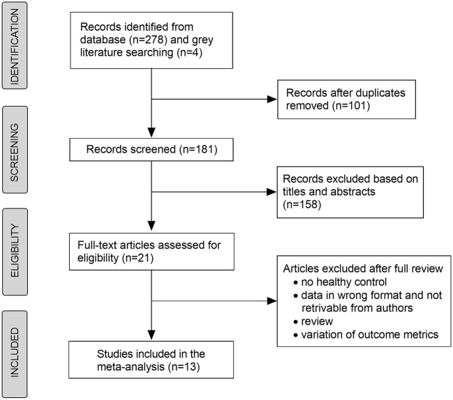
PubMed and Google Scholar were searched for studies that compared retinal and choroidal microvasculature between MS and healthy controls (HC) with OCTA. MS patients were divided into 2 groups: MS with (MSON) or without optic neuritis (MSNON).

RESULTS

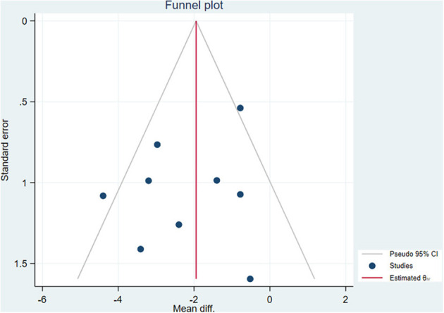
Totally, 13 studies including 996 MS eyes and 847 HC eyes were included. Compared with the HC, the vessel density of the whole superficial vascular complex (SVC) was reduced by 2.27% and 4.30% in the MSNON and MSON groups, respectively. The peripapillary vessel density was 2.28% lower and 4.96% lower in the MSNON and MSON groups, respectively, than in the HC. Furthermore, the MSON group had significant lower vessel density of the SVC (mean difference [MD] = -2.17%, < 0.01) and lower peripapillary vessel density (MD = -2.02%, = 0.02) than the MSNON group. No significant difference was found in the deep vascular complex or choriocapillaris densities among MSNON, MSON or HC groups ( > 0.05). Meta-regression analyses suggested that illness duration and the Expanded Disability Status Scale scores of MS patients were possible sources of heterogeneity ( < 0.05).

CONCLUSION

The retinal SVC and peripapillary vessel density decreased significantly in MS eyes, especially in eyes with optic neuritis. Retinal microvasculature is a potential biomarker of disease progression in MS.

摘要

目的

进行一项系统评价和荟萃分析,以利用光学相干断层扫描血管造影(OCTA)研究多发性硬化症(MS)患者视网膜和脉络膜微血管的变化。

方法

在PubMed和谷歌学术上搜索使用OCTA比较MS患者与健康对照(HC)之间视网膜和脉络膜微血管的研究。MS患者分为两组:患有视神经炎的MS(MSON)和未患有视神经炎的MS(MSNON)。

结果

总共纳入了13项研究,包括996只MS患者眼和847只HC眼。与HC相比,MSNON组和MSON组整个浅表血管复合体(SVC)的血管密度分别降低了2.27%和4.30%。MSNON组和MSON组视乳头周围血管密度分别比HC组低2.28%和4.96%。此外,MSON组的SVC血管密度(平均差[MD]=-2.17%,<0.01)和视乳头周围血管密度(MD=-2.02%,=0.02)显著低于MSNON组。在MSNON组、MSON组或HC组之间,深部血管复合体或脉络膜毛细血管密度未发现显著差异(>0.05)。荟萃回归分析表明,MS患者的病程和扩展残疾状态量表评分可能是异质性的来源(<0.05)。

结论

MS患者眼中视网膜SVC和视乳头周围血管密度显著降低,尤其是在患有视神经炎的眼中。视网膜微血管是MS疾病进展的潜在生物标志物。

https://cdn.ncbi.nlm.nih.gov/pmc/blobs/9e70/9880267/3fc427a18ee5/fnins-16-1121899-g0002.jpg
https://cdn.ncbi.nlm.nih.gov/pmc/blobs/9e70/9880267/8956e33970a1/fnins-16-1121899-g0001.jpg
https://cdn.ncbi.nlm.nih.gov/pmc/blobs/9e70/9880267/3fc427a18ee5/fnins-16-1121899-g0002.jpg
https://cdn.ncbi.nlm.nih.gov/pmc/blobs/9e70/9880267/8956e33970a1/fnins-16-1121899-g0001.jpg
https://cdn.ncbi.nlm.nih.gov/pmc/blobs/9e70/9880267/3fc427a18ee5/fnins-16-1121899-g0002.jpg

相似文献

1
Microvascular impairments detected by optical coherence tomography angiography in multiple sclerosis patients: A systematic review and meta-analysis.光学相干断层扫描血管造影术检测多发性硬化症患者的微血管损伤:一项系统评价和荟萃分析。
Front Neurosci. 2023 Jan 13;16:1121899. doi: 10.3389/fnins.2022.1121899. eCollection 2022.
2
Optical coherence tomography angiography measurements in multiple sclerosis: a systematic review and meta-analysis.光学相干断层扫描血管造影测量多发性硬化症:系统评价和荟萃分析。
J Neuroinflammation. 2023 Mar 27;20(1):85. doi: 10.1186/s12974-023-02763-4.
3
Retinal layer segmentation in multiple sclerosis: a systematic review and meta-analysis.多发性硬化症中的视网膜层分割:系统评价和荟萃分析。
Lancet Neurol. 2017 Oct;16(10):797-812. doi: 10.1016/S1474-4422(17)30278-8. Epub 2017 Sep 12.
4
Significant retinal microvascular impairments in multiple sclerosis assessed through optical coherence tomography angiography.通过光学相干断层扫描血管造影评估多发性硬化症患者显著的视网膜微血管损伤。
Mult Scler Relat Disord. 2023 Feb;70:104505. doi: 10.1016/j.msard.2023.104505. Epub 2023 Jan 4.
5
Retinal optical coherence tomography measures in multiple sclerosis: a systematic review and meta-analysis.多发性硬化症的视网膜光学相干断层扫描测量:系统评价和荟萃分析。
Ann Clin Transl Neurol. 2024 Sep;11(9):2236-2253. doi: 10.1002/acn3.52165. Epub 2024 Jul 28.
6
Shortened Pattern Electroretinogram Latency and Impaired Autoregulatory Dynamics to Steady-State Stimuli in Patients With Multiple Sclerosis.多发性硬化症患者短潜伏期图形视网膜电图和对稳态刺激的自动调节动力学受损。
J Neuroophthalmol. 2021 Mar 1;41(1):60-68. doi: 10.1097/WNO.0000000000000894.
7
Visual Function and Disability Are Associated with Increased Retinal Volumetric Vessel Density in Patients with Multiple Sclerosis.多发性硬化症患者的视觉功能和残疾与视网膜容积血管密度增加相关。
Am J Ophthalmol. 2020 May;213:34-45. doi: 10.1016/j.ajo.2019.12.021. Epub 2020 Jan 8.
8
The effect of optic neuritis attacks on choroidal vascularity index in patients with multiple sclerosis.多发性硬化症患者视神经炎发作对脉络膜血流指数的影响。
Graefes Arch Clin Exp Ophthalmol. 2021 Aug;259(8):2413-2424. doi: 10.1007/s00417-021-05143-x. Epub 2021 Mar 25.
9
Alterations in the retinal vasculature occur in multiple sclerosis and exhibit novel correlations with disability and visual function measures.视网膜血管的改变发生在多发性硬化症中,并与残疾和视觉功能测量具有新的相关性。
Mult Scler. 2020 Jun;26(7):815-828. doi: 10.1177/1352458519845116. Epub 2019 May 16.
10
Microvascular blood flow velocities measured with a retinal function imager: inter-eye correlations in healthy controls and an exploration in multiple sclerosis.使用视网膜功能成像仪测量的微血管血流速度:健康对照者的双眼相关性及在多发性硬化症中的探索
Eye Vis (Lond). 2018 Nov 2;5:29. doi: 10.1186/s40662-018-0123-0. eCollection 2018.

引用本文的文献

1
Optical coherence tomography and angiography reveal early retinal alterations in pediatric-onset multiple sclerosis.光学相干断层扫描和血管造影显示儿童期多发性硬化症患者早期视网膜改变。
Eur J Pediatr. 2025 Jun 6;184(7):398. doi: 10.1007/s00431-025-06234-1.
2
Barriers in Healthcare to the Use of Optical Coherence Tomography Angiography in Multiple Sclerosis.医疗保健领域中多发性硬化症使用光学相干断层扫描血管造影术的障碍。
Neurol Ther. 2025 Feb;14(1):45-56. doi: 10.1007/s40120-024-00670-1. Epub 2024 Nov 5.
3
Oculomics analysis in multiple sclerosis: Current ophthalmic clinical and imaging biomarkers.

本文引用的文献

1
Retinal and Choriocapillary Vascular Changes in Early Stages of Multiple Sclerosis: A Prospective Study.多发性硬化早期视网膜和脉络膜毛细血管血管变化:一项前瞻性研究。
J Clin Med. 2021 Dec 9;10(24):5756. doi: 10.3390/jcm10245756.
2
Retinal Oxygen Metabolism and Haemodynamics in Patients With Multiple Sclerosis and History of Optic Neuritis.患有多发性硬化症和视神经炎病史患者的视网膜氧代谢与血流动力学
Front Neurosci. 2021 Oct 12;15:761654. doi: 10.3389/fnins.2021.761654. eCollection 2021.
3
Optical coherence tomography angiography indicates subclinical retinal disease in neuromyelitis optica spectrum disorders.
眼科学分析在多发性硬化症中的应用:当前的眼科临床和影像学生物标志物。
Eye (Lond). 2024 Oct;38(14):2701-2710. doi: 10.1038/s41433-024-03132-y. Epub 2024 Jun 10.
4
Non-Invasive Evaluation of Retinal Vascular Alterations in a Mouse Model of Optic Neuritis Using Laser Speckle Flowgraphy and Optical Coherence Tomography Angiography.采用激光散斑血流成像和光相干断层扫描血管造影术对实验性变态反应性视神经炎模型小鼠视网膜血管改变的无创评估。
Cells. 2023 Nov 22;12(23):2685. doi: 10.3390/cells12232685.
5
Retina and microvascular alterations in migraine: a systemic review and meta-analysis.偏头痛中的视网膜和微血管改变:一项系统评价与荟萃分析。
Front Neurol. 2023 Sep 28;14:1241778. doi: 10.3389/fneur.2023.1241778. eCollection 2023.
光学相干断层扫描血管造影显示视神经脊髓炎谱系疾病的亚临床视网膜病变。
Mult Scler. 2022 Apr;28(4):522-531. doi: 10.1177/13524585211028831. Epub 2021 Jul 14.
4
Macular vessel density differs in multiple sclerosis and neuromyelitis optica spectrum disorder: An optical coherence tomography angiography study.多发性硬化症和视神经脊髓炎谱系障碍的黄斑血管密度不同:一项光学相干断层扫描血管造影研究。
PLoS One. 2021 Jun 17;16(6):e0253417. doi: 10.1371/journal.pone.0253417. eCollection 2021.
5
Evaluation of optical coherence tomography angiography findings in patients with multiple sclerosis.多发性硬化症患者光学相干断层扫描血管造影检查结果的评估。
Indian J Ophthalmol. 2021 Jun;69(6):1457-1463. doi: 10.4103/ijo.IJO_2964_20.
6
Optical Coherence Tomography Angiography of Peripapillary Vessel Density in Multiple Sclerosis and Neuromyelitis Optica Spectrum Disorder: A Comparative Study.多发性硬化症和视神经脊髓炎谱系障碍中视乳头周围血管密度的光学相干断层扫描血管造影:一项比较研究
J Clin Med. 2021 Feb 5;10(4):609. doi: 10.3390/jcm10040609.
7
Optical Coherence Tomography Angiography (OCTA) in Multiple Sclerosis and Neuromyelitis Optica Spectrum Disorder.光学相干断层扫描血管造影术(OCTA)在多发性硬化症和视神经脊髓炎谱系障碍中的应用
Front Neurol. 2020 Dec 10;11:604049. doi: 10.3389/fneur.2020.604049. eCollection 2020.
8
Angiography with optical coherence tomography as a biomarker in multiple sclerosis.光学相干断层扫描血管造影作为多发性硬化症的生物标志物。
PLoS One. 2020 Dec 8;15(12):e0243236. doi: 10.1371/journal.pone.0243236. eCollection 2020.
9
Retinal changes in Alzheimer's disease- integrated prospects of imaging, functional and molecular advances.阿尔茨海默病的视网膜变化——影像学、功能和分子进展的综合展望。
Prog Retin Eye Res. 2021 May;82:100899. doi: 10.1016/j.preteyeres.2020.100899. Epub 2020 Sep 2.
10
Optical coherence tomography angiography in multiple sclerosis: A cross-sectional study.光学相干断层扫描血管造影在多发性硬化症中的应用:一项横断面研究。
PLoS One. 2020 Jul 23;15(7):e0236090. doi: 10.1371/journal.pone.0236090. eCollection 2020.