Cicione Claudia, Vadalà Gianluca, Di Giacomo Giuseppina, Tilotta Veronica, Ambrosio Luca, Russo Fabrizio, Zampogna Biagio, Cannata Francesca, Papalia Rocco, Denaro Vincenzo
Laboratory for Regenerative Orthopaedics, Research Unit of Orthopaedic and Trauma Surgery, Department of Medicine and Surgery, Università Campus Bio-Medico di Roma, Rome, Italy.
Operative Research Unit of Orthopaedic and Trauma Surgery, Fondazione Policlinico Universitario Campus Bio-Medico, Rome, Italy.
Front Bioeng Biotechnol. 2023 Jan 17;11:911600. doi: 10.3389/fbioe.2023.911600. eCollection 2023.
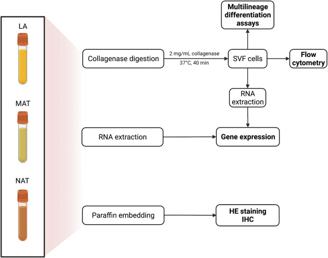
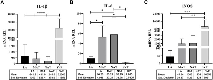
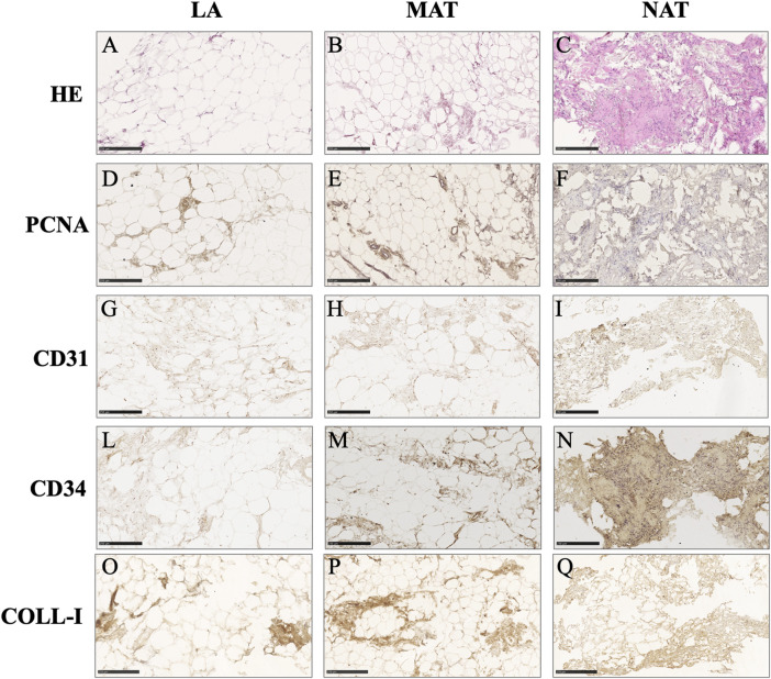
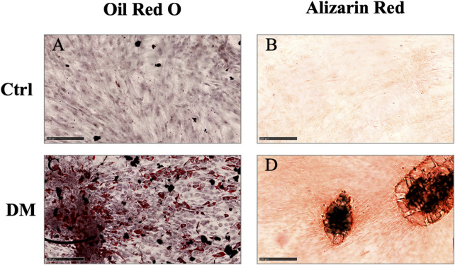
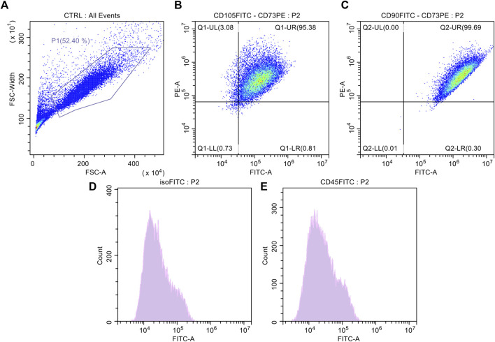
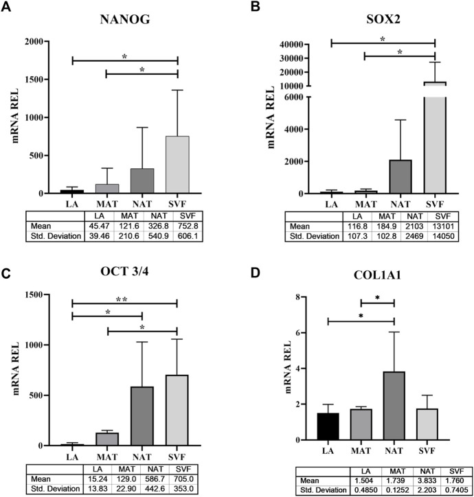
Adipose tissue is widely exploited in regenerative medicine thanks to its trophic properties, mainly based on the presence of adipose-derived stromal cells. Numerous devices have been developed to promote its clinical use, leading to the introduction of one-step surgical procedures to obtain minimally manipulated adipose tissue derivatives. However, only a few studies compared their biological properties. This study aimed to characterize micro-fragmented (MAT) and nanofat adipose tissue (NAT) obtained with two different techniques. MAT, NAT and unprocessed lipoaspirate were collected from surgical specimens. RNA extraction and collagenase isolation of stromal vascular fraction (SVF) were performed. Tissue sections were analysed by histological and immunohistochemical (collagen type I, CD31, CD34 and PCNA) staining to assess tissue morphology and cell content. qPCR was performed to evaluate the expression of stemness-related (, and ), extracellular matrix () and inflammatory genes ( and ). Furthermore, multilineage differentiation was assessed following culture in adipogenic and osteogenic media and staining with Oil Red O and Alizarin red. ASC immunophenotype was assessed by flow cytometric analysis of CD90, CD105, CD73 and CD45. Histological and immunohistochemical results showed an increased amount of stroma and a reduction of adipocytes in MAT and NAT, with the latter displaying the highest content of collagen type I, CD31, CD34 and PCNA. From LA to MAT and NAT, an increasing expression of , , , and was noted, while no significant differences in terms of and emerged. No statistically significant differences were noted between NAT and SVF in terms of stemness-related genes, while the latter demonstrated a significantly higher expression of stress-related markers. SVF cells derived from all three samples (LA, MAT, and NAT) showed a similar ASC immunoprofile as well as osteogenic and adipogenic differentiation. Our results showed that both MAT and NAT techniques allowed the rapid isolation of ASC-rich grafts with a high anabolic and proliferative potential. However, NAT showed the highest levels of extracellular matrix content, replicating cells, and stemness gene expression. These results may provide precious clues for the use of adipose tissue derivatives in the clinical setting.
由于其营养特性,脂肪组织在再生医学中得到了广泛应用,这主要基于脂肪来源的基质细胞的存在。已经开发了许多装置来促进其临床应用,从而引入了一步外科手术程序以获得最少操作的脂肪组织衍生物。然而,只有少数研究比较了它们的生物学特性。本研究旨在表征通过两种不同技术获得的微片段化脂肪组织(MAT)和纳米脂肪组织(NAT)。从手术标本中收集MAT、NAT和未处理的抽脂物。进行基质血管成分(SVF)的RNA提取和胶原酶分离。通过组织学和免疫组织化学(I型胶原、CD31、CD34和增殖细胞核抗原)染色分析组织切片,以评估组织形态和细胞含量。进行qPCR以评估干性相关基因(、和)、细胞外基质()和炎症基因(和)的表达。此外,在成脂和成骨培养基中培养并用油红O和茜素红染色后评估多谱系分化。通过对CD90、CD105、CD73和CD45的流式细胞术分析评估ASC免疫表型。组织学和免疫组织化学结果显示,MAT和NAT中的基质数量增加,脂肪细胞减少,后者显示I型胶原、CD31、CD34和增殖细胞核抗原的含量最高。从抽脂物到MAT和NAT,观察到、、、和的表达增加,而和方面没有显著差异。在干性相关基因方面,NAT和SVF之间没有统计学上的显著差异,而后者显示应激相关标志物的表达显著更高。来自所有三个样本(抽脂物、MAT和NAT)的SVF细胞显示出相似的ASC免疫谱以及成骨和成脂分化。我们的结果表明,MAT和NAT技术都能快速分离出富含ASC且具有高合成代谢和增殖潜力的移植物。然而,NAT显示出最高水平的细胞外基质含量、复制细胞和干性基因表达。这些结果可能为脂肪组织衍生物在临床环境中的应用提供宝贵线索。