Department of Molecular Biology and Genetics, Krishna Institute of Medical Sciences 'Deemed to be University', Taluka-Karad, Dist-Satara, Pin, 415 539, Maharashtra, India.
Pharm Nanotechnol. 2023 Jun 6;11(3):303-314. doi: 10.2174/2211738511666230206112537.
Recent advancements in biomedicine have revolutionized nanomedicine as a therapeutic moderator in the management of both infectious and noninfectious diseases.
In the current study we demonstrated biosynthesis of gold nanoparticles using aqueous leaf extract of as a capping and reducing agent and evaluation of their antioxidant, antibacterial, and anticancer properties.
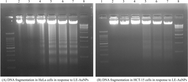
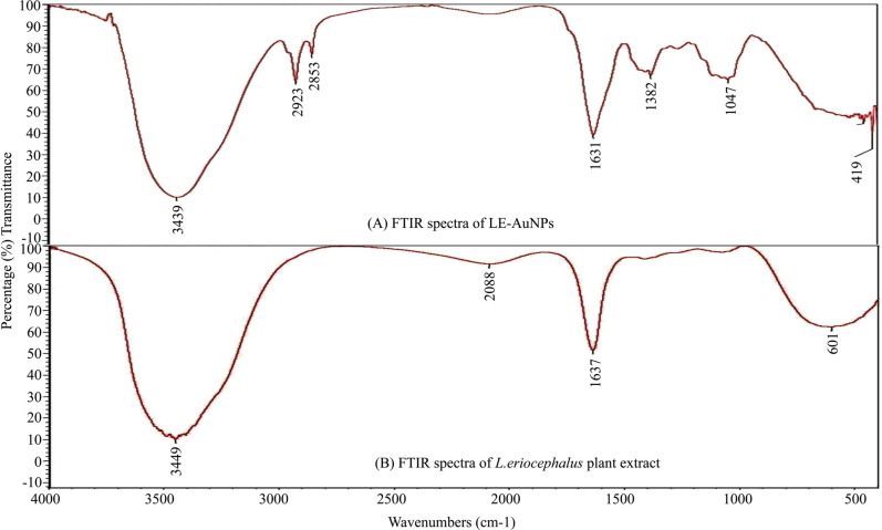
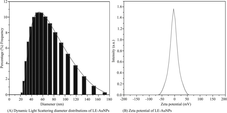
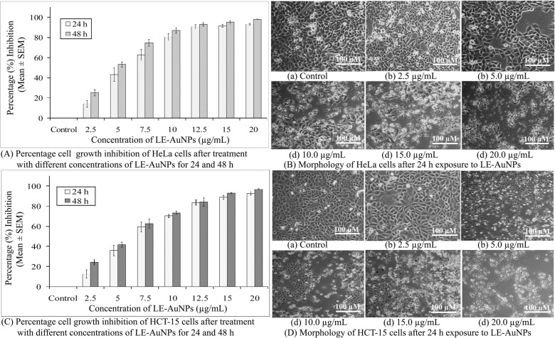
The biosynthesized LE-AuNPs were characterized by UV-Vis spectrophotometry, SEM, TEM, XRD, FTIR, DLS, and Zeta potential analysis. The antibacterial activity was checked by a minimum inhibitory concentration assay. The anticancer potential of biogenic LE-AuNPs was checked by cytotoxicity and genotoxicity assay against HeLa and HCT-15 cells.
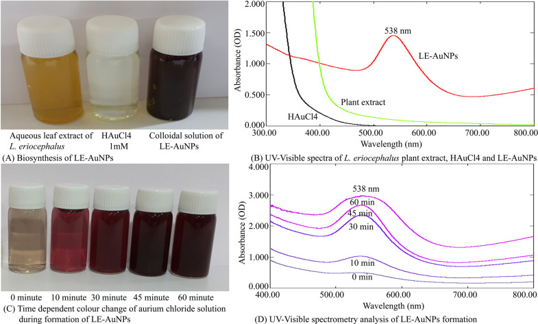
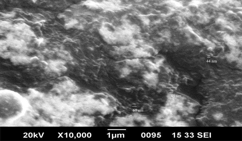
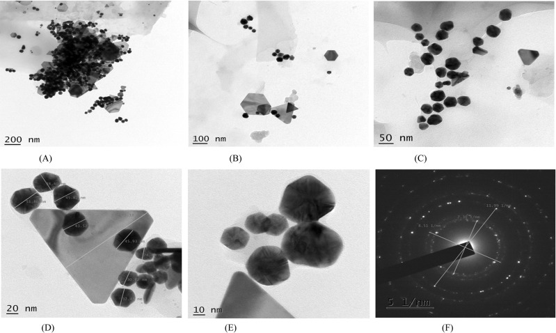
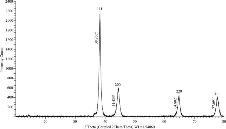
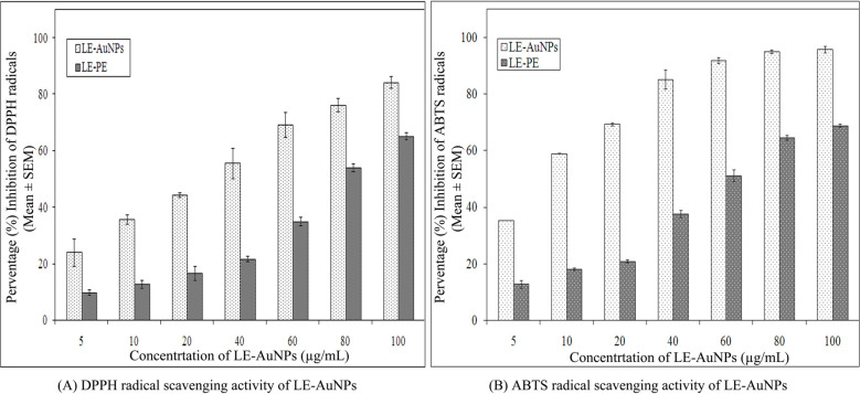
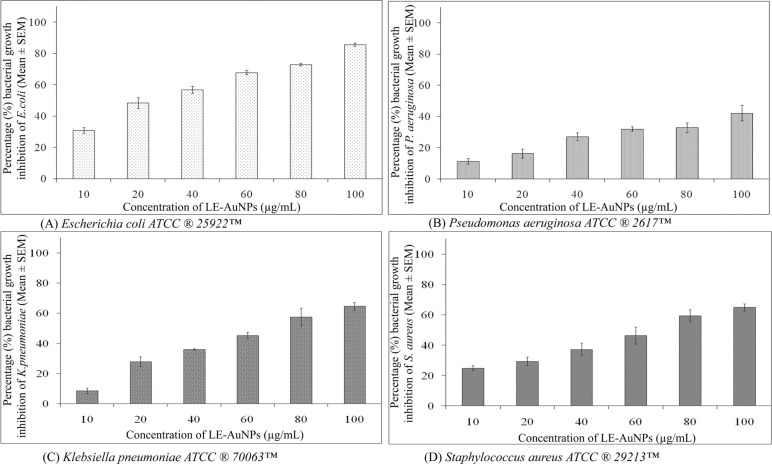
The characteristic surface plasmon resonance peak of the colloidal solution at 538 nm by UV-Vis spectrum confirmed the formation of LE-AuNPs in the solution. The SEM, TEM, and XRD revealed 20-60 sized hexagonal and crystalline LE-AuNPs. The LE-AuNPs displayed significant inhibition potential against DPPH and ABTS radicals in vitro. The LE-AuNPs demonstrated significant antibacterial potential. The results of cytotoxicity interpreted that biogenic gold nanoparticles exhibited strong dose and time-dependent cytotoxicity effect against selected cancer cell lines where IC50 of LE-AuNPs required to inhibit the growth of HeLa cells after 24 h and 48 h exposure were 5.65± 0.69 μg/mL and 4.37±0.23 μg/mL respectively and that of HCT- 15 cells was 6.46 ± 0.69 μg/mL and 5.27 ± 0.34 μg/mL, 24h and 48h post-exposure respectively.
Findings from this study revealed that gold nanoparticles synthesized using showed remarkable antioxidant, antimicrobial, and extensive cytotoxicity and genotoxicity activities.
最近生物医学的进步使纳米医学成为治疗传染性和非传染性疾病的治疗调节剂。
本研究旨在利用作为封端和还原剂,通过水提树叶法合成金纳米粒子,并评估其抗氧化、抗菌和抗癌特性。
通过紫外-可见分光光度法、SEM、TEM、XRD、FTIR、DLS 和 Zeta 电位分析对生物合成的 LE-AuNPs 进行了表征。通过最低抑菌浓度测定法检查了抗菌活性。通过细胞毒性和遗传毒性试验,检查了生物合成的 LE-AuNPs 对 HeLa 和 HCT-15 细胞的抗癌潜力。
紫外-可见光谱的胶体溶液特征表面等离子体共振峰在 538nm 处证实了溶液中 LE-AuNPs 的形成。SEM、TEM 和 XRD 显示 20-60 纳米大小的六边形和结晶 LE-AuNPs。LE-AuNPs 在体外对 DPPH 和 ABTS 自由基具有显著的抑制潜力。LE-AuNPs 表现出显著的抗菌潜力。细胞毒性试验结果表明,生物合成的金纳米粒子对选定的癌细胞系具有很强的剂量和时间依赖性细胞毒性作用,LE-AuNPs 抑制 HeLa 细胞生长的 IC50 在 24h 和 48h 暴露后分别为 5.65±0.69μg/ml 和 4.37±0.23μg/ml,HCT-15 细胞的 IC50 分别为 6.46±0.69μg/ml 和 5.27±0.34μg/ml,暴露后 24h 和 48h。
本研究结果表明,利用 合成的金纳米粒子具有显著的抗氧化、抗菌和广泛的细胞毒性和遗传毒性活性。