Yeo Miji, Yoon Jung Won, Park Gyu Tae, Shin Sung-Chan, Song Young-Cheol, Cheon Yong-Il, Lee Byung-Joo, Kim Geun Hyung, Kim Jae Ho
Department of Biomechatronic Engineering, College of Biotechnology and Bioengineering, Sungkyunkwan University (SKKU), Suwon 16419, Republic of Korea.
Department of Engineering Science and Mechanics, Penn State University, University Park, PA 16802, USA.
Mater Today Bio. 2023 Jan 26;19:100564. doi: 10.1016/j.mtbio.2023.100564. eCollection 2023 Apr.
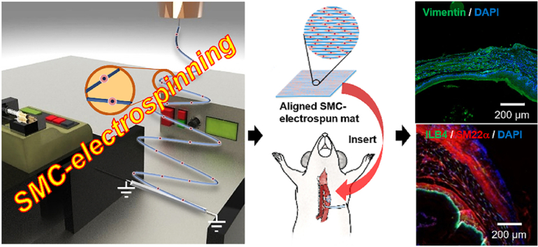
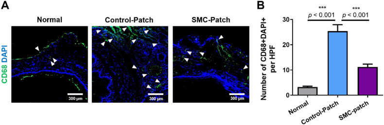
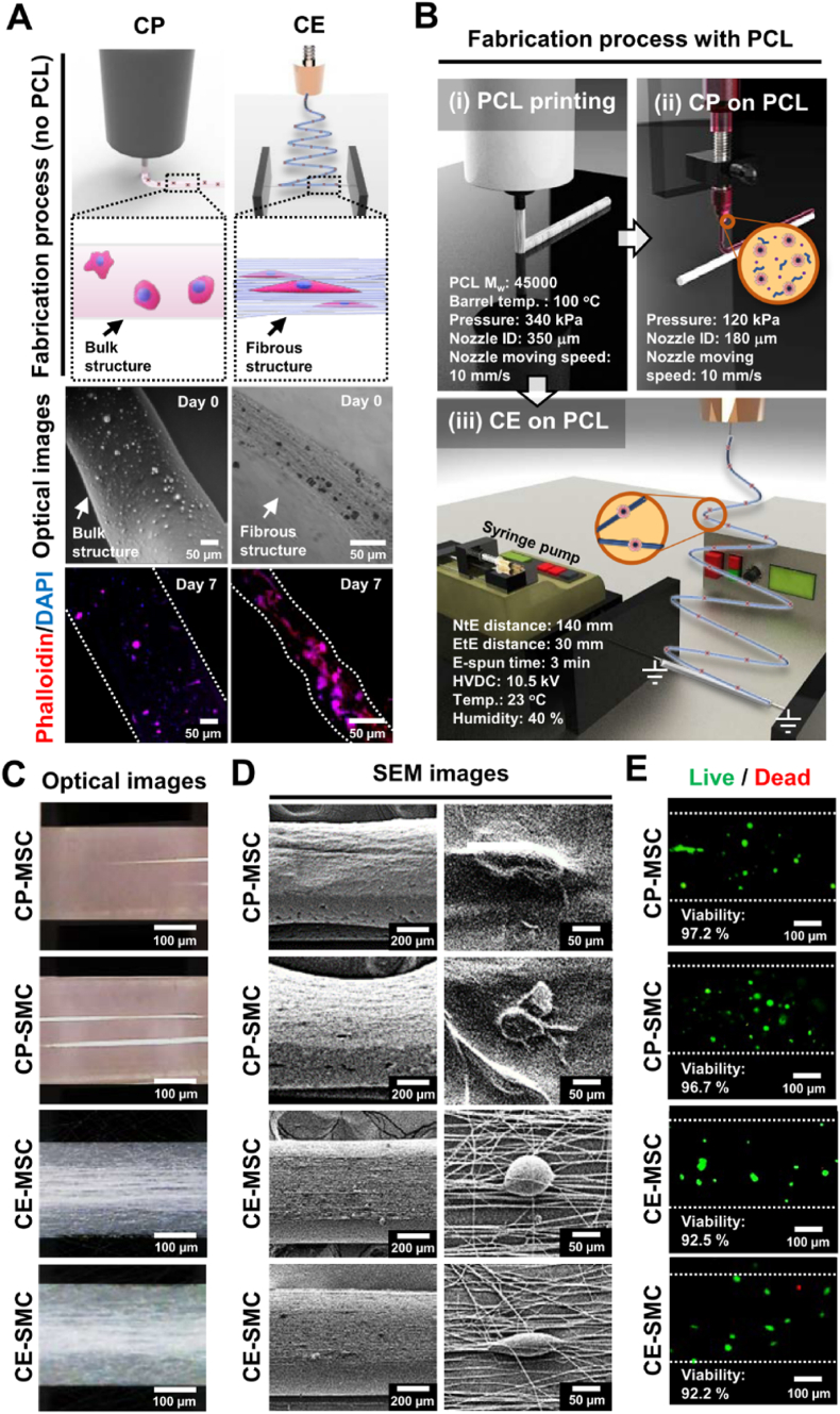
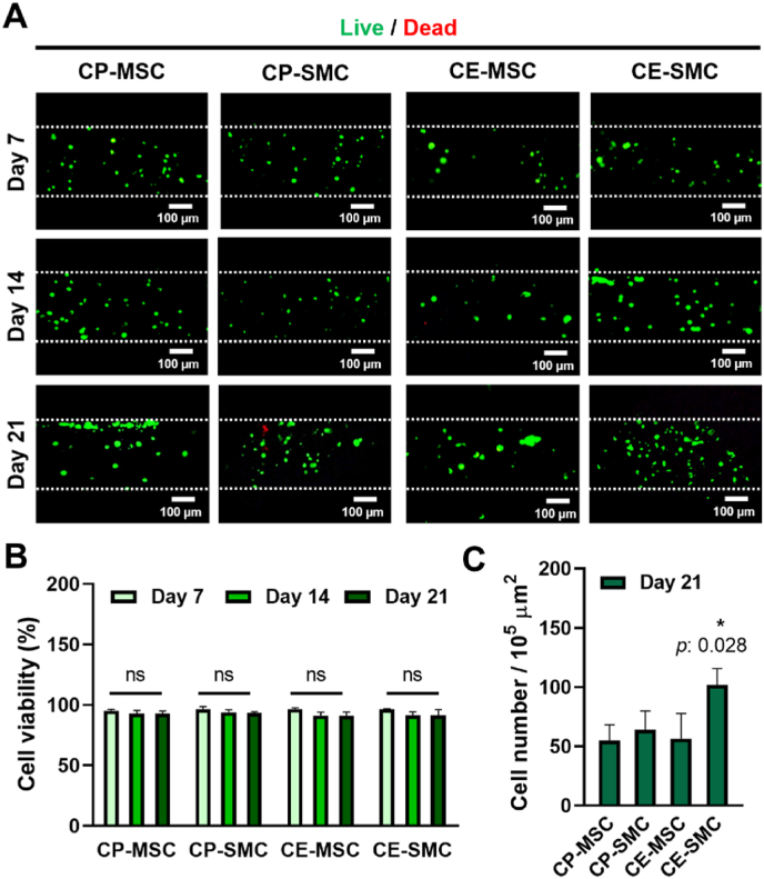
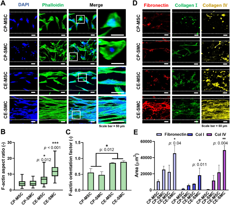
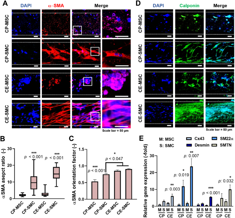
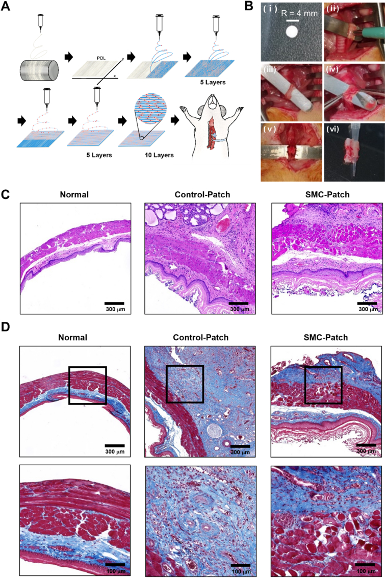
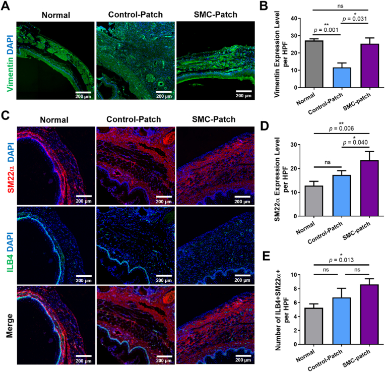
The esophagus exhibits peristalsis via contraction of circularly and longitudinally aligned smooth muscles, and esophageal replacement is required if there is a critical-sized wound. In this study, we proposed to reconstruct esophageal tissues using cell electrospinning (CE), an advanced technique for encapsulating living cells into fibers that allows control of the direction of fiber deposition. After treatment with transforming growth factor-β, mesenchymal stem cell-derived smooth muscle cells (SMCs) were utilized for cell electrospinning or three-dimensional bioprinting to compare the effects of aligned micropatterns on cell morphology. CE resulted in SMCs with uniaxially arranged and elongated cell morphology with upregulated expression levels of SMC-specific markers, including connexin 43, smooth muscle protein 22 alpha (SM22α), desmin, and smoothelin. When SMC-laden nanofibrous patches were transplanted into a rat esophageal defect model, the SMC patch promoted regeneration of esophageal wounds with an increased number of newly formed blood vessels and enhanced the SMC-specific markers of SM22α and vimentin. Taken together, CE with its advantages, such as guidance of highly elongated, aligned cell morphology and accelerated SMC differentiation, can be an efficient strategy to reconstruct smooth muscle tissues and treat esophageal perforation.
食管通过环形和纵向排列的平滑肌收缩表现出蠕动,若存在临界尺寸的伤口,则需要进行食管置换。在本研究中,我们提议使用细胞静电纺丝(CE)来重建食管组织,这是一种将活细胞封装到纤维中以控制纤维沉积方向的先进技术。在用转化生长因子-β处理后,利用间充质干细胞衍生的平滑肌细胞(SMC)进行细胞静电纺丝或三维生物打印,以比较排列微图案对细胞形态的影响。CE导致SMC呈现单轴排列且拉长的细胞形态,平滑肌特异性标志物的表达水平上调,包括连接蛋白43、平滑肌蛋白22α(SM22α)、结蛋白和平滑肌肌动蛋白。当将负载SMC的纳米纤维贴片移植到大鼠食管缺损模型中时,SMC贴片促进了食管伤口的再生,新生血管数量增加,并增强了SM22α和波形蛋白的SMC特异性标志物。综上所述,CE具有引导高度拉长、排列的细胞形态以及加速SMC分化等优点,可成为重建平滑肌组织和治疗食管穿孔的有效策略。