Department of Neurosurgery, Jena University Hospital - Friedrich Schiller University Jena, Jena, Germany.
Department for Radiology, Jena University Hospital - Friedrich Schiller University Jena, Jena, Germany.
J Orthop Surg Res. 2023 Feb 10;18(1):93. doi: 10.1186/s13018-023-03560-8.
The assessment of bone density is of great importance nowadays due to the increasing age of patients. Especially in regard to the surgical stabilization of the spine, the assessment of bone density is important for therapeutic decision making. The aim of this work was to record trabecular bone density values using Hounsfield units of the second cervical vertebra.
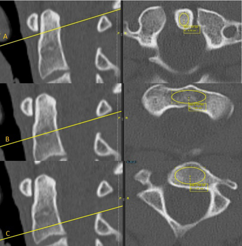
The study is a monocentric retrospective data analysis of 198 patients who received contrast-enhanced polytrauma computed tomography in a period of two years at a maximum care hospital. Hounsfield units were measured in three different regions within the C2: dens, transition area between dens and vertebral body and vertebral body. The measured Hounsfield units were converted into bone density values using a validated formula.
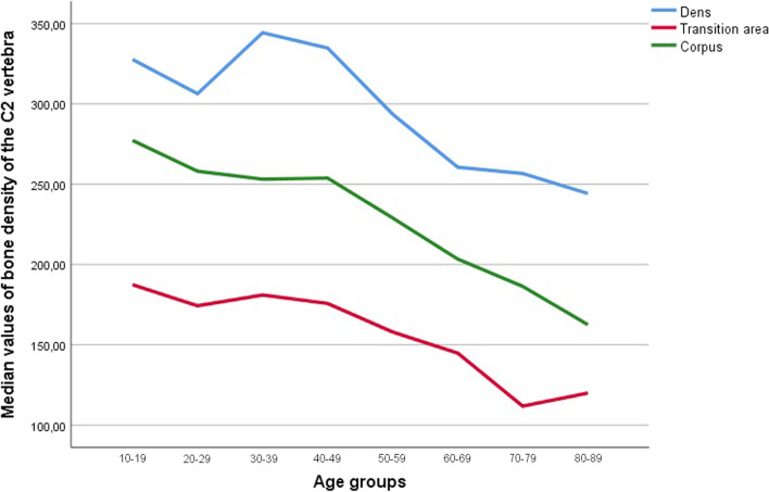
A total of 198 patients were included. The median bone density varied in different regions of all measured C2 vertebrae: in the dens axis, C2 transition area between dens and vertebral body, and in the vertebral body bone densities were 302.79 mg/cm, 160.08 mg/cm, and 240.31 mg/cm, respectively. The transition area from dens axis to corpus had statistically significant lower bone density values compared to the other regions (p < 0.001). There was a decrease in bone density values after age 50 years in both men and women (p < 0.001).
The transitional area from dens axis to corpus showed statistically significant lower bone density values compared to the adjacent regions (p < 0.001). This area seems to be a predilection site for fractures of the 2nd cervical vertebra, which is why special attention should be paid here in radiological diagnostics after a trauma.
由于患者年龄的增长,骨密度的评估变得越来越重要。特别是在脊柱的外科稳定中,骨密度的评估对于治疗决策非常重要。本研究的目的是使用第二颈椎的亨氏单位记录小梁骨密度值。
这是一项在一家重症监护医院在两年时间内对 198 名接受增强型多发伤计算机断层扫描的患者进行的单中心回顾性数据分析。在 C2 内的三个不同区域测量了亨氏单位:齿状突、齿状突与椎体之间的过渡区和椎体。使用经过验证的公式将测量的亨氏单位转换为骨密度值。
共纳入 198 例患者。所有测量的 C2 椎体的不同区域的骨密度中位数存在差异:在齿状突轴、齿状突与椎体之间的 C2 过渡区以及椎体的骨密度分别为 302.79 mg/cm、160.08 mg/cm 和 240.31 mg/cm。与其他区域相比,齿状突轴到椎体的过渡区的骨密度值具有统计学显著差异(p < 0.001)。男性和女性的骨密度值均在 50 岁后下降(p < 0.001)。
与相邻区域相比,齿状突轴到椎体的过渡区的骨密度值具有统计学显著差异(p < 0.001)。该区域似乎是第 2 颈椎骨折的易患部位,因此在创伤后放射学诊断中应特别注意该区域。