Balomenos Dimitrios B, Gouletsou Pagona G, Galatos Apostolos D
Clinic of Surgery, Faculty of Veterinary Science, School of Health Sciences, University of Thessaly, 43100 Karditsa, Greece.
Clinic of Obstetrics and Reproduction, Faculty of Veterinary Science, School of Health Sciences, University of Thessaly, Trikalon 224, 43100 Karditsa, Greece.
Animals (Basel). 2023 Jan 27;13(3):426. doi: 10.3390/ani13030426.
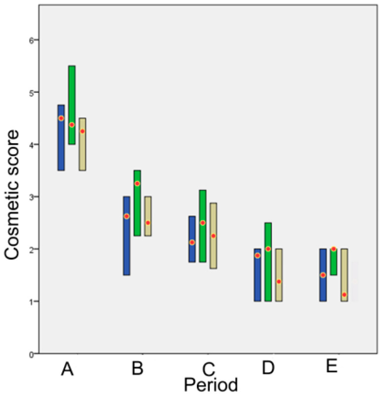
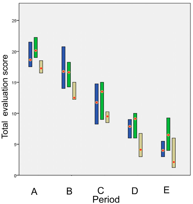
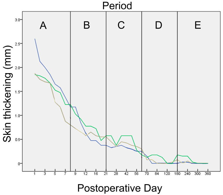
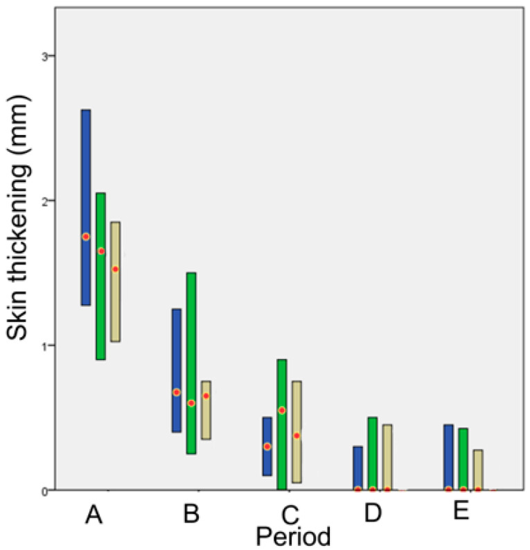
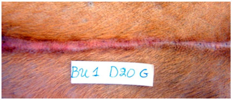
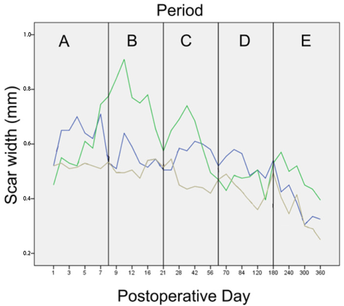
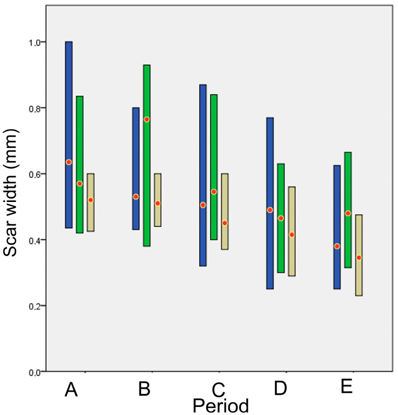
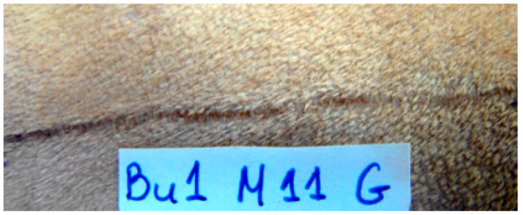
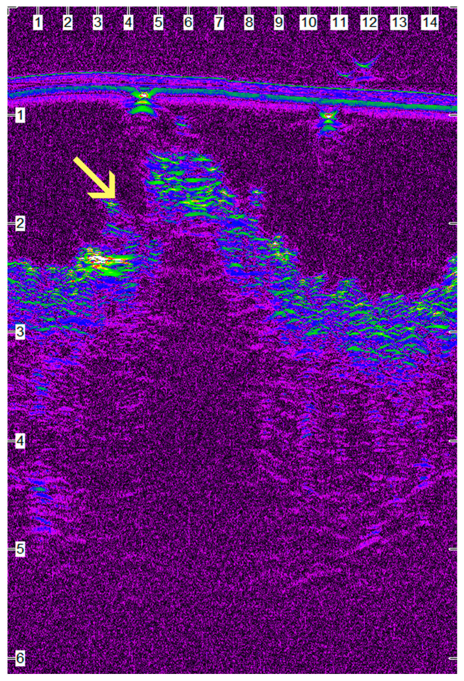
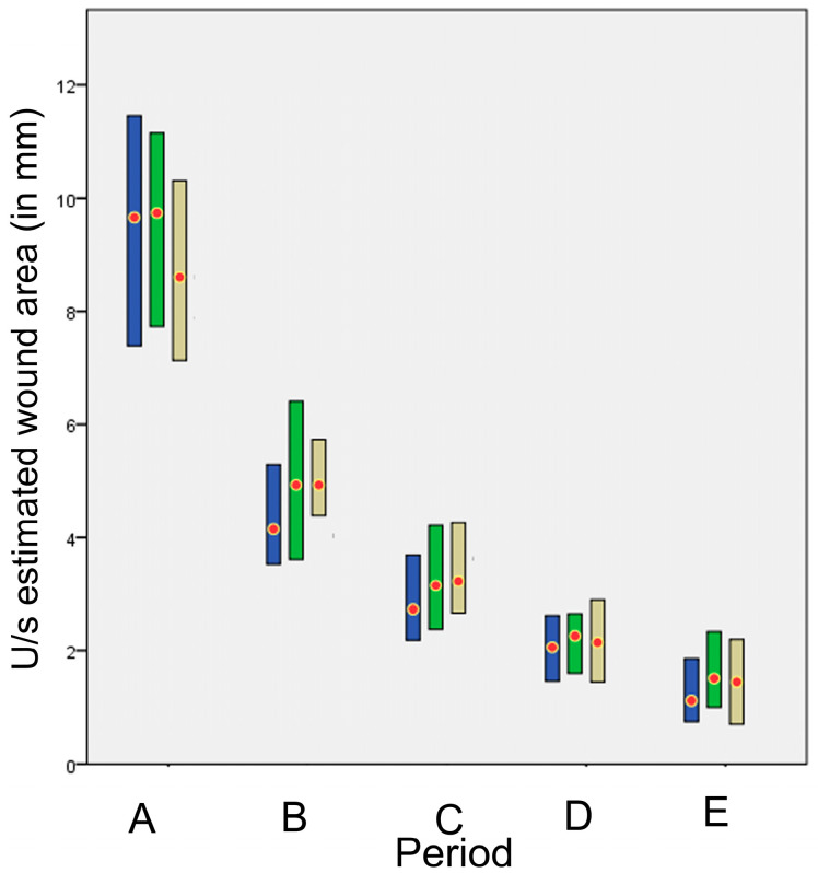
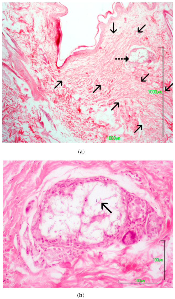
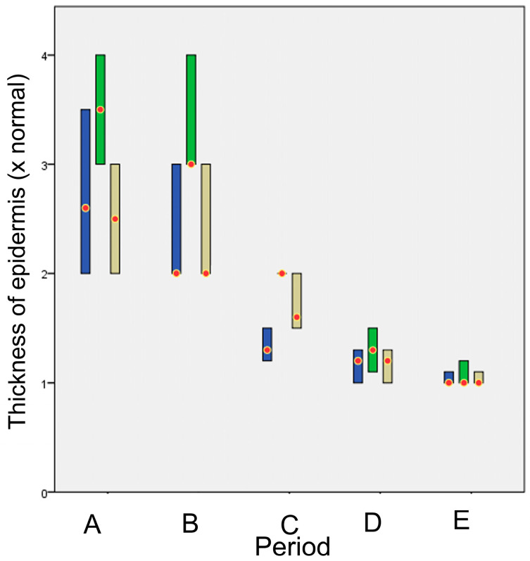
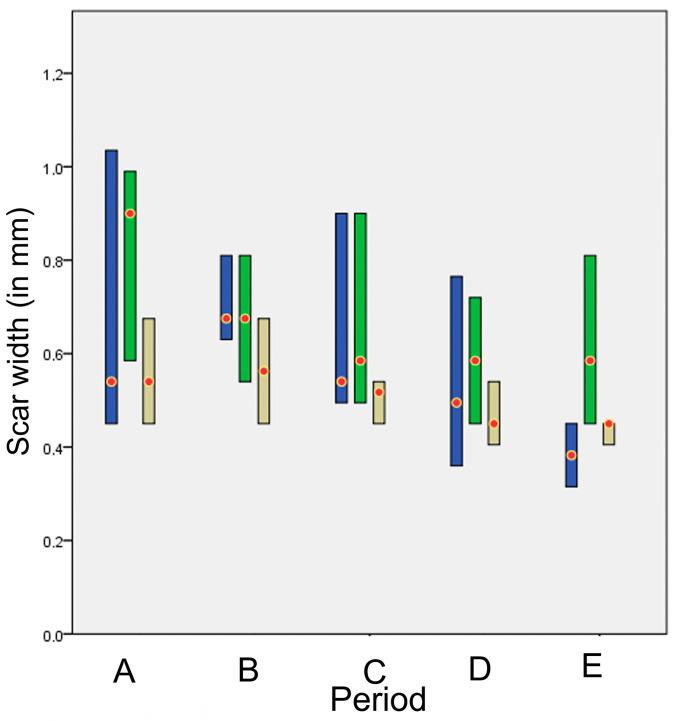
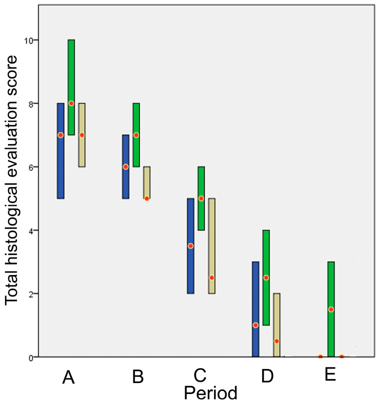
The study aimed to monitor the healing process in the canine skin following surgical incision and closure using staples or tissue glue and to compare them with the intradermal suture pattern. Surgically created skin incisions in 10 dogs were apposed with staples, tissue glue (n-butyl cyanoacrylate) and continuous intradermal pattern. The cosmetic appearance of the wounds was blindly evaluated on days 7, 14 and 28 and once a month until the end of the experiment, i.e., one year after the incision. Ultrasonographic and clinical evaluation was performed on days 0-10, 12, 14, 16, 18, 21, 24 and 28, once a week until the end of the 3rd month and once a month until the end of the experiment. Histopathological evaluation was performed on days 7, 14, 28, 180 and 365. The median time required for the performance of each technique differed significantly between techniques; stapling lasted 21 s, glue 2 min 16 s and intradermal 15 min 37 s. Cosmetic appearance with glue was statistically worse than staples and intradermal. The clinical appearance of intradermal was significantly better than glue and staples. No significant differences were found at histological evaluation; however, glue had the worst score throughout the experiment. The overall evaluation of the techniques showed that glue had the worst score compared to intradermal and staples, with the difference being statistically significant in the first postoperative week. Intradermal suture pattern is much better than glue application for skin closure in dogs, whilst is not significantly better than staples. Staples should be preferred when time is an important factor.
该研究旨在监测犬类皮肤在使用吻合钉或组织胶水进行手术切口和闭合后的愈合过程,并将其与皮内缝合模式进行比较。在10只犬身上 surgically created皮肤切口,分别采用吻合钉、组织胶水(正丁基氰基丙烯酸酯)和连续皮内缝合模式进行对位缝合。在第7天、14天和28天以及直至实验结束(即切口后一年)每月对伤口的外观进行盲法评估。在第0 - 10天、12天、14天、16天、18天、21天、24天和28天进行超声检查和临床评估,在第3个月结束前每周进行一次,直至实验结束每月进行一次。在第7天、14天、28天、180天和365天进行组织病理学评估。每种技术操作所需的中位时间在不同技术之间存在显著差异;吻合钉操作持续21秒,胶水操作持续2分16秒,皮内缝合操作持续15分37秒。使用胶水时的外观在统计学上比吻合钉和皮内缝合差。皮内缝合的临床外观明显优于胶水和吻合钉。在组织学评估中未发现显著差异;然而,在整个实验过程中胶水的评分最差。对这些技术的总体评估表明,与皮内缝合和吻合钉相比,胶水的评分最差,在术后第一周差异具有统计学意义。对于犬类皮肤闭合,皮内缝合模式比使用胶水要好得多,虽然不比吻合钉显著更好。当时间是一个重要因素时,应优先选择吻合钉。 (注:surgically created 这里可能表述有误,推测可能是“手术造成的”之类意思,但不确定准确表述)