• 文献检索
  • 文档翻译
  • 深度研究
  • 学术资讯
  • Suppr Zotero 插件Zotero 插件
  • 邀请有礼
  • 套餐&价格
  • 历史记录
应用&插件
Suppr Zotero 插件Zotero 插件浏览器插件Mac 客户端Windows 客户端微信小程序
定价
高级版会员购买积分包购买API积分包
服务
文献检索文档翻译深度研究API 文档MCP 服务
关于我们
关于 Suppr公司介绍联系我们用户协议隐私条款
关注我们

Suppr 超能文献

核心技术专利:CN118964589B侵权必究
粤ICP备2023148730 号-1Suppr @ 2026

文献检索

告别复杂PubMed语法,用中文像聊天一样搜索,搜遍4000万医学文献。AI智能推荐,让科研检索更轻松。

立即免费搜索

文件翻译

保留排版,准确专业,支持PDF/Word/PPT等文件格式,支持 12+语言互译。

免费翻译文档

深度研究

AI帮你快速写综述,25分钟生成高质量综述,智能提取关键信息,辅助科研写作。

立即免费体验

深度学习模型在子宫腺肌病超声诊断中的应用。

Application of Deep Learning Model in the Sonographic Diagnosis of Uterine Adenomyosis.

机构信息

Division of Gynecology and Human Reproduction Physiopathology, IRCCS Azienda Ospedaliero-Universitaria di Bologna, 40126 Bologna, Italy.

Department of Medical and Surgical Sciences (DIMEC), University of Bologna, 40126 Bologna, Italy.

出版信息

Int J Environ Res Public Health. 2023 Jan 18;20(3):1724. doi: 10.3390/ijerph20031724.

DOI:10.3390/ijerph20031724
PMID:36767092
原文链接:https://pmc.ncbi.nlm.nih.gov/articles/PMC9914280/
Abstract

BACKGROUND

This study aims to evaluate the diagnostic performance of Deep Learning (DL) machine for the detection of adenomyosis on uterine ultrasonographic images and compare it to intermediate ultrasound skilled trainees.

METHODS

Prospective observational study were conducted between 1 and 30 April 2022. Transvaginal ultrasound (TVUS) diagnosis of adenomyosis was investigated by an experienced sonographer on 100 fertile-age patients. Videoclips of the uterine corpus were recorded and sequential ultrasound images were extracted. Intermediate ultrasound-skilled trainees and DL machine were asked to make a diagnosis reviewing uterine images. We evaluated and compared the accuracy, sensitivity, positive predictive value, F1-score, specificity and negative predictive value of the DL model and the trainees for adenomyosis diagnosis.

RESULTS

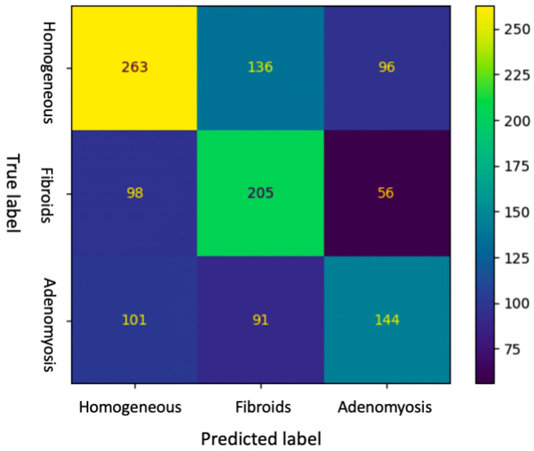
Accuracy of DL and intermediate ultrasound-skilled trainees for the diagnosis of adenomyosis were 0.51 (95% CI, 0.48-0.54) and 0.70 (95% CI, 0.60-0.79), respectively. Sensitivity, specificity and F1-score of DL were 0.43 (95% CI, 0.38-0.48), 0.82 (95% CI, 0.79-0.85) and 0.46 (0.42-0.50), respectively, whereas intermediate ultrasound-skilled trainees had sensitivity of 0.72 (95% CI, 0.52-0.86), specificity of 0.69 (95% CI, 0.58-0.79) and F1-score of 0.55 (95% CI, 0.43-0.66).

CONCLUSIONS

In this preliminary study DL model showed a lower accuracy but a higher specificity in diagnosing adenomyosis on ultrasonographic images compared to intermediate-skilled trainees.

摘要

背景

本研究旨在评估深度学习(DL)机器在经阴道超声图像上检测子宫腺肌病的诊断性能,并将其与中级超声熟练医师进行比较。

方法

前瞻性观察性研究于 2022 年 4 月 1 日至 30 日进行。由一位有经验的超声医师对 100 名生育期患者进行经阴道超声(TVUS)诊断子宫腺肌病。记录子宫体的视频片段并提取连续的超声图像。中级超声熟练医师和 DL 机器被要求通过查看子宫图像做出诊断。我们评估并比较了 DL 模型和中级超声熟练医师对腺肌病诊断的准确性、敏感度、阳性预测值、F1 评分、特异性和阴性预测值。

结果

DL 和中级超声熟练医师诊断子宫腺肌病的准确率分别为 0.51(95%CI,0.48-0.54)和 0.70(95%CI,0.60-0.79)。DL 的敏感度、特异性和 F1 评分分别为 0.43(95%CI,0.38-0.48)、0.82(95%CI,0.79-0.85)和 0.46(0.42-0.50),而中级超声熟练医师的敏感度为 0.72(95%CI,0.52-0.86)、特异性为 0.69(95%CI,0.58-0.79)和 F1 评分为 0.55(95%CI,0.43-0.66)。

结论

在这项初步研究中,与中级熟练医师相比,DL 模型在超声图像上诊断子宫腺肌病的准确性较低,但特异性较高。

https://cdn.ncbi.nlm.nih.gov/pmc/blobs/8718/9914280/cd3b700f19a5/ijerph-20-01724-g001.jpg

相似文献

1
Application of Deep Learning Model in the Sonographic Diagnosis of Uterine Adenomyosis.深度学习模型在子宫腺肌病超声诊断中的应用。
Int J Environ Res Public Health. 2023 Jan 18;20(3):1724. doi: 10.3390/ijerph20031724.
2
Question Mark Sign and Transvaginal Ultrasound Uterine Tenderness for the Diagnosis of Adenomyosis: A Prospective Validation.问号征及经阴道超声子宫压痛对子宫腺肌病的诊断:一项前瞻性验证研究
J Ultrasound Med. 2020 Jul;39(7):1405-1412. doi: 10.1002/jum.15237. Epub 2020 Feb 7.
3
Ultrasound diagnosis of endometriosis and adenomyosis: State of the art.子宫内膜异位症和子宫腺肌病的超声诊断:现状。
Best Pract Res Clin Obstet Gynaecol. 2018 Aug;51:16-24. doi: 10.1016/j.bpobgyn.2018.01.013. Epub 2018 Feb 14.
4
Transvaginal sonographic criteria for the diagnosis of adenomyosis based on histopathologic correlation.经阴道超声诊断标准基于组织病理学相关性的子宫腺肌病。
Taiwan J Obstet Gynecol. 2010 Mar;49(1):40-4. doi: 10.1016/S1028-4559(10)60007-1.
5
Comparison of Sensitivity and Specificity of Structured and Narrative Reports of Transvaginal Ultrasonogaphy for Adenomyosis.经阴道超声检查中结构性报告与叙事性报告对子宫腺肌病的敏感性和特异性比较。
J Minim Invasive Gynecol. 2021 Jun;28(6):1216-1224. doi: 10.1016/j.jmig.2020.11.001. Epub 2020 Nov 15.
6
Sonographic Assessment of Uterine Biometry for the Diagnosis of Diffuse Adenomyosis in a Tertiary Outpatient Clinic.三级门诊中经超声测量子宫生物指标诊断弥漫性子宫腺肌病的研究
J Pers Med. 2022 Sep 23;12(10):1572. doi: 10.3390/jpm12101572.
7
Accuracy of findings in the diagnosis of uterine adenomyosis on ultrasound.超声诊断子宫腺肌病的准确性。
Abdom Radiol (NY). 2020 Mar;45(3):842-850. doi: 10.1007/s00261-019-02231-9.
8
Accuracy of ultrasound signs on two-dimensional transvaginal ultrasound in prediction of adenomyosis: prospective multicenter study.二维经阴道超声声像图征象对子宫腺肌病的预测准确性:前瞻性多中心研究。
Ultrasound Obstet Gynecol. 2023 Nov;62(5):739-746. doi: 10.1002/uog.26197.
9
Diagnostic Accuracy of Transvaginal Ultrasound in Adenomyosis Taking MRI as a Gold Standard.经 MRI 作为金标准诊断的经阴道超声在子宫腺肌病中的诊断准确性。
J Coll Physicians Surg Pak. 2023 Oct;33(10):1118-1123. doi: 10.29271/jcpsp.2023.10.1118.
10
A Detailed Study in Adenomyosis and Endometriosis: Evaluation of the Rate of Coexistence Between Uterine Adenomyosis and DIE According to Imaging and Histopathology Findings.子宫腺肌病和子宫内膜异位症的详细研究:根据影像学和组织病理学结果评估子宫腺肌病和深部浸润型子宫内膜异位症共存的发生率。
Reprod Sci. 2021 Aug;28(8):2387-2397. doi: 10.1007/s43032-021-00527-0. Epub 2021 Mar 16.

引用本文的文献

1
Perspectives in transvaginal sonography for the diagnosis of adenomyosis.经阴道超声检查在子宫腺肌病诊断中的应用前景。
Front Med (Lausanne). 2025 Jun 19;12:1533648. doi: 10.3389/fmed.2025.1533648. eCollection 2025.
2
Risk factors for uterine adenomyosis diagnosed by MRI in women of reproductive age.育龄期女性中通过磁共振成像(MRI)诊断子宫腺肌病的风险因素。
Ann Med Surg (Lond). 2025 Mar 9;87(4):1941-1946. doi: 10.1097/MS9.0000000000003175. eCollection 2025 Apr.
3
Artificial intelligence applications in endometriosis imaging.人工智能在子宫内膜异位症成像中的应用。

本文引用的文献

1
Consensus on revised definitions of Morphological Uterus Sonographic Assessment (MUSA) features of adenomyosis: results of modified Delphi procedure.关于子宫腺肌病形态学超声评估(MUSA)特征修订定义的共识:改良 Delphi 程序的结果。
Ultrasound Obstet Gynecol. 2022 Jul;60(1):118-131. doi: 10.1002/uog.24786.
2
Uterine Fibroids and Infertility.子宫肌瘤与不孕
Diagnostics (Basel). 2021 Aug 12;11(8):1455. doi: 10.3390/diagnostics11081455.
3
Application of artificial intelligence in gynecologic malignancies: A review.人工智能在妇科恶性肿瘤中的应用:综述
Abdom Radiol (NY). 2025 Apr 1. doi: 10.1007/s00261-025-04897-w.
4
Application of artificial intelligence to ultrasound imaging for benign gynecological disorders: systematic review.人工智能在良性妇科疾病超声成像中的应用:系统评价
Ultrasound Obstet Gynecol. 2025 Mar;65(3):295-302. doi: 10.1002/uog.29171. Epub 2025 Jan 31.
5
How Reproducible Are the Ultrasound Features of Adenomyosis Defined by the Revised MUSA Consensus?经修订的MUSA共识所定义的子宫腺肌病超声特征的可重复性如何?
J Clin Med. 2025 Jan 13;14(2):456. doi: 10.3390/jcm14020456.
6
Achieving enhanced diagnostic precision in endometrial lesion analysis through a data enhancement framework.通过数据增强框架提高子宫内膜病变分析中的诊断精度。
Front Oncol. 2024 Oct 15;14:1440881. doi: 10.3389/fonc.2024.1440881. eCollection 2024.
7
A stacked machine learning-based classification model for endometriosis and adenomyosis: a retrospective cohort study utilizing peripheral blood and coagulation markers.一种基于堆叠机器学习的子宫内膜异位症和子宫腺肌病分类模型:一项利用外周血和凝血标志物的回顾性队列研究。
Front Digit Health. 2024 Sep 10;6:1463419. doi: 10.3389/fdgth.2024.1463419. eCollection 2024.
8
From Diagnosis to Fertility: Optimizing Treatment of Adenomyosis for Reproductive Health.从诊断到生育:优化子宫腺肌病治疗以促进生殖健康
J Clin Med. 2024 Aug 21;13(16):4926. doi: 10.3390/jcm13164926.
9
Prediction of adenomyosis according to revised definitions of morphological uterus sonographic assessment features.根据形态学子宫超声评估特征的修订定义预测子宫腺肌病。
Front Med (Lausanne). 2024 Aug 8;11:1387515. doi: 10.3389/fmed.2024.1387515. eCollection 2024.
10
Deep learning based uterine fibroid detection in ultrasound images.基于深度学习的超声图像子宫肌瘤检测。
BMC Med Imaging. 2024 Aug 19;24(1):218. doi: 10.1186/s12880-024-01389-z.
J Obstet Gynaecol Res. 2021 Aug;47(8):2577-2585. doi: 10.1111/jog.14818. Epub 2021 May 10.
4
Artificial intelligence (AI) in the detection of rectosigmoid deep endometriosis.人工智能(AI)在直肠乙状结肠深部子宫内膜异位症检测中的应用。
Eur J Obstet Gynecol Reprod Biol. 2021 Jun;261:29-33. doi: 10.1016/j.ejogrb.2021.04.012. Epub 2021 Apr 14.
5
Diagnostic Accuracy of Transvaginal Ultrasound and Magnetic Resonance Imaging for Adenomyosis: Systematic Review and Meta-Analysis and Review of Sonographic Diagnostic Criteria.经阴道超声与磁共振成像诊断子宫腺肌病的准确性:系统评价与荟萃分析,以及超声诊断标准综述。
J Ultrasound Med. 2021 Nov;40(11):2289-2306. doi: 10.1002/jum.15635. Epub 2021 Jan 27.
6
Ultrasound image analysis using deep neural networks for discriminating between benign and malignant ovarian tumors: comparison with expert subjective assessment.基于深度神经网络的超声图像分析用于鉴别良恶性卵巢肿瘤:与专家主观评估的比较。
Ultrasound Obstet Gynecol. 2021 Jan;57(1):155-163. doi: 10.1002/uog.23530.
7
Early Learning Curve in the Assessment of Deep Pelvic Endometriosis for Ultrasound and Magnetic Resonance Imaging.超声和磁共振成像评估深部盆腔子宫内膜异位症的早期学习曲线。
Biomed Res Int. 2020 Sep 26;2020:8757281. doi: 10.1155/2020/8757281. eCollection 2020.
8
Question Mark Sign and Transvaginal Ultrasound Uterine Tenderness for the Diagnosis of Adenomyosis: A Prospective Validation.问号征及经阴道超声子宫压痛对子宫腺肌病的诊断:一项前瞻性验证研究
J Ultrasound Med. 2020 Jul;39(7):1405-1412. doi: 10.1002/jum.15237. Epub 2020 Feb 7.
9
Noninvasive Diagnosis of Adenomyosis: A Structured Review and Meta-analysis of Diagnostic Accuracy in Imaging.影像学对子宫腺肌病的无创性诊断:系统评价和诊断准确性的 Meta 分析。
J Minim Invasive Gynecol. 2020 Feb;27(2):408-418.e3. doi: 10.1016/j.jmig.2019.11.001. Epub 2019 Nov 8.
10
New Sonographic Classification of Adenomyosis: Do Type and Degree of Adenomyosis Correlate to Severity of Symptoms?腺肌病的新超声分类:腺肌病的类型和程度与症状严重程度相关吗?
J Minim Invasive Gynecol. 2020 Sep-Oct;27(6):1308-1315. doi: 10.1016/j.jmig.2019.09.788. Epub 2019 Oct 7.