• 文献检索
  • 文档翻译
  • 深度研究
  • 学术资讯
  • Suppr Zotero 插件Zotero 插件
  • 邀请有礼
  • 套餐&价格
  • 历史记录
应用&插件
Suppr Zotero 插件Zotero 插件浏览器插件Mac 客户端Windows 客户端微信小程序
定价
高级版会员购买积分包购买API积分包
服务
文献检索文档翻译深度研究API 文档MCP 服务
关于我们
关于 Suppr公司介绍联系我们用户协议隐私条款
关注我们

Suppr 超能文献

核心技术专利:CN118964589B侵权必究
粤ICP备2023148730 号-1Suppr @ 2026

文献检索

告别复杂PubMed语法,用中文像聊天一样搜索,搜遍4000万医学文献。AI智能推荐,让科研检索更轻松。

立即免费搜索

文件翻译

保留排版,准确专业,支持PDF/Word/PPT等文件格式,支持 12+语言互译。

免费翻译文档

深度研究

AI帮你快速写综述,25分钟生成高质量综述,智能提取关键信息,辅助科研写作。

立即免费体验

新冠病毒康复者视网膜的光学相干断层扫描血管造影参数

Optical Coherence Tomography Angiography Parameters of the Retina in SARS-CoV-2 Recovered Subjects.

作者信息

Sodhi Punita K, Arora Ritu, Kumar Suresh, Jaisingh Kirti, R Archana T, Rao Kavya C, Chhabra Karan, Saxena Sonal, Manchanda Vikas, Sharma Shantanu

机构信息

Ophthalmology, Guru Nanak Eye Centre and Maulana Azad Medical College, New Delhi, IND.

Medicine, Maulana Azad Medical College, New Delhi, IND.

出版信息

Cureus. 2023 Jan 9;15(1):e33548. doi: 10.7759/cureus.33548. eCollection 2023 Jan.

DOI:10.7759/cureus.33548
PMID:36779162
原文链接:https://pmc.ncbi.nlm.nih.gov/articles/PMC9907863/
Abstract

INTRODUCTION

This study aims to evaluate retinochoroidal optical coherence tomography angiography (OCTA) parameters in patients recovered from severe acute respiratory syndrome coronavirus 2 (SARS-CoV-2).

METHODS

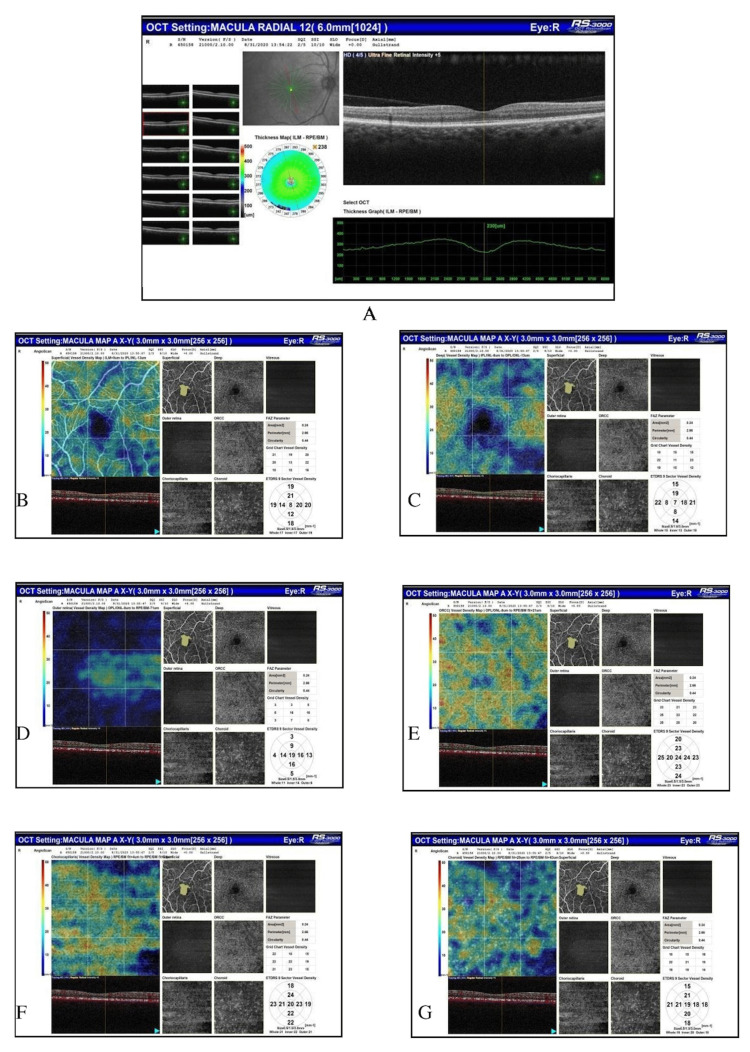
This study was an observational study that included 80 subjects being discharged after having negative reports on the reverse transcription-polymerase chain reaction (RT-PCR) test for SARS-CoV-2 to evaluate OCTA parameters of the retina. The subjects underwent an ophthalmic evaluation that included best-corrected visual acuity (BCVA), intraocular pressure (IOP), color vision (CV), contrast sensitivity (CS), and optical coherence tomography (OCT) parameters. OCTA was done for all patients and was evaluated for foveal avascular zone (FAZ) area, perimeter, and circularity index, and vessel density (VD) in superficial capillary plexus (SCP), deep capillary plexus (DCP), outer retina (OR), outer retina chorio-capillaries (ORCC), chorio-capillaries (CC), and choroid (C) using 3 x 3 mm scans. The OCTA parameters were compared with normative data of the Indian population for various parameters in question.

RESULTS

The subjects included 54/80 (67.5%) males and 26/80 (32.5%) females having a mean age of 52.40 ± 15.71 (18-60) years. The systemic evaluation revealed 38.75% of subjects had hypertension, 30% had diabetes, 20% had kidney disease, 5% had tuberculosis, and 3.75% had coronary artery disease. The mean distance BCVA was logarithm of the minimum angle of resolution (LogMAR) (1.17 ± 0.22), mean IOP was 17.0 ± 4.0 mmHg, mean CS was 2.13 ± 0.36, 50.62% of subjects had normal CV on Farnsworth test while 47% had tritanopia, and none of the subjects had red-green CV defect on Ishihara plates. The OCT scan was normal in 90% of eyes while the posterior vitreous detachment was seen in 4% of eyes, broad vitreomacular adhesion in 2.5% of eyes, and the globally adherent epiretinal membrane was seen in 2.5% of eyes. The mean central macular thickness (CMT) measured 245.14 ± 28.41 micrometers. The mean FAZ area measured 0.37 ± 0.15 mm, the perimeter was 3.28 ± 1.08 mm, and the circularity index measured 0.41 ± 0.10. The average VD in SCP measured 16.06 ± 12.29, in DCP measured 9.11 ± 8.75, in OR measured 6.38 ± 7.37, in ORCC measured 42.53 ± 12.46, in CC measured 25.83 ± 16.31, and in C measured 25.52 ± 17.49. The VD in coronavirus disease 2019 (COVID-19) subjects was significantly lesser than that in the healthy Indian population in all layers except ORCC.

CONCLUSIONS

The SARS-CoV-2 recovered subjects have a reduced VD in retinochoroidal layers from COVID-19, an underlying systemic disease, or both. The CS values fall within normal limits. Several subjects show tritanopia on the Farnsworth test but no red-green CV defect on Ishihara plates.

摘要

引言

本研究旨在评估从严重急性呼吸综合征冠状病毒2(SARS-CoV-2)感染中康复的患者的视网膜脉络膜光学相干断层扫描血管造影(OCTA)参数。

方法

本研究为一项观察性研究,纳入了80名严重急性呼吸综合征冠状病毒2逆转录聚合酶链反应(RT-PCR)检测呈阴性后出院的受试者,以评估其视网膜的OCTA参数。受试者接受了眼科评估,包括最佳矫正视力(BCVA)、眼压(IOP)、色觉(CV)、对比敏感度(CS)和光学相干断层扫描(OCT)参数。所有患者均进行了OCTA检查,并使用3×3mm扫描评估了黄斑无血管区(FAZ)面积、周长和圆形度指数,以及浅表毛细血管丛(SCP)、深层毛细血管丛(DCP)、外层视网膜(OR)、外层视网膜脉络膜毛细血管(ORCC)、脉络膜毛细血管(CC)和脉络膜(C)的血管密度(VD)。将OCTA参数与印度人群相关参数的正常数据进行比较。

结果

受试者包括54/80(67.5%)名男性和26/80(32.5%)名女性,平均年龄为52.40±15.71(18 - 60)岁。全身评估显示,38.75%的受试者患有高血压,30%患有糖尿病,20%患有肾脏疾病,5%患有结核病,3.75%患有冠状动脉疾病。平均BCVA为最小分辨角对数(LogMAR)(1.17±0.22),平均IOP为17.0±4.0mmHg,平均CS为2.13±0.36,50.62%的受试者在Farnsworth测试中色觉正常,47%患有蓝色色盲,且在石原氏色盲测试中无一受试者存在红绿色觉缺陷。90%的眼睛OCT扫描正常,4%的眼睛可见玻璃体后脱离,2.5%的眼睛可见广泛的玻璃体黄斑粘连,2.5%的眼睛可见全层附着的视网膜前膜。平均中心黄斑厚度(CMT)为245.14±28.41微米。平均FAZ面积为0.37±0.15mm,周长为3.28±1.08mm,圆形度指数为0.41±0.10。SCP的平均VD为16.06±12.29,DCP的平均VD为9.11±8.75,OR的平均VD为6.38±7.37,ORCC的平均VD为42.53±12.46,CC的平均VD为25.83±16.31,C的平均VD为25.52±17.49。2019冠状病毒病(COVID-19)受试者各层的VD均显著低于健康印度人群,外层视网膜脉络膜毛细血管层(ORCC)除外。

结论

从SARS-CoV-2感染中康复的受试者,其视网膜脉络膜各层的VD因COVID-19、潜在的全身性疾病或两者共同作用而降低。CS值在正常范围内。部分受试者在Farnsworth测试中表现为蓝色色盲,但在石原氏色盲测试中无红绿色觉缺陷。

https://cdn.ncbi.nlm.nih.gov/pmc/blobs/8662/9907863/eb172dcdaa8f/cureus-0015-00000033548-i01.jpg

相似文献

1
Optical Coherence Tomography Angiography Parameters of the Retina in SARS-CoV-2 Recovered Subjects.新冠病毒康复者视网膜的光学相干断层扫描血管造影参数
Cureus. 2023 Jan 9;15(1):e33548. doi: 10.7759/cureus.33548. eCollection 2023 Jan.
2
Evaluating the Quantitative Foveal Avascular Zone and Retino-Choroidal Vessel Density Using Optical Coherence Tomography Angiography in a Healthy Indian Population.在健康印度人群中使用光学相干断层扫描血管造影术评估定量黄斑无血管区和视网膜脉络膜血管密度
Cureus. 2022 Aug 4;14(8):e27669. doi: 10.7759/cureus.27669. eCollection 2022 Aug.
3
Optical Coherence Tomography Angiography Parameters in Indian Patients With Central Serous Chorioretinopathy.印度中心性浆液性脉络膜视网膜病变患者的光学相干断层扫描血管造影参数
Cureus. 2023 Oct 4;15(10):e46467. doi: 10.7759/cureus.46467. eCollection 2023 Oct.
4
The Association of Neovascularization With Optical Coherence Tomography Angiography Parameters in Proliferative Diabetic Retinopathy.增殖性糖尿病视网膜病变中新生血管形成与光学相干断层扫描血管造影参数的相关性
Cureus. 2023 May 29;15(5):e39633. doi: 10.7759/cureus.39633. eCollection 2023 May.
5
[Correlation of capillary plexus with visual acuity in idiopathic macular epiretinal membrane eyes using optical coherence tomography angiography].[利用光学相干断层扫描血管造影术研究特发性黄斑视网膜前膜眼中毛细血管丛与视力的相关性]
Zhonghua Yan Ke Za Zhi. 2019 Oct 11;55(10):757-762. doi: 10.3760/cma.j.issn.0412-4081.2019.10.006.
6
Assessment of macular microvasculature features before and after vitrectomy in the idiopathic macular epiretinal membrane using a grading system: An optical coherence tomography angiography study.采用分级系统评估特发性黄斑视网膜前膜玻璃体切除术前、后黄斑微血管特征:光学相干断层扫描血管造影研究。
Acta Ophthalmol. 2021 Nov;99(7):e1168-e1175. doi: 10.1111/aos.14753. Epub 2021 Jan 10.
7
Correlations Between Optical Coherence Tomography Angiography Parameters and the Visual Acuity in Patients with Diabetic Retinopathy.糖尿病视网膜病变患者光学相干断层扫描血管造影参数与视力的相关性
Clin Ophthalmol. 2020 Apr 23;14:1107-1115. doi: 10.2147/OPTH.S248881. eCollection 2020.
8
Foveal avascular zone and macular vessel density after correction for magnification error in unilateral amblyopia using optical coherence tomography angiography.应用光学相干断层扫描血管造影术矫正单侧弱视的放大误差后黄斑中心凹无血管区和黄斑血管密度。
BMC Ophthalmol. 2019 Aug 5;19(1):171. doi: 10.1186/s12886-019-1177-z.
9
Optical coherence tomography angiography analysis of the retina in patients recovered from COVID-19: a case-control study.COVID-19康复患者视网膜的光学相干断层扫描血管造影分析:一项病例对照研究。
Can J Ophthalmol. 2021 Feb;56(1):24-30. doi: 10.1016/j.jcjo.2020.11.006. Epub 2020 Nov 14.
10
Deep Capillary Plexus as Biomarker of Peripheral Capillary Nonperfusion in Central Retinal Vein Occlusion.深层毛细血管丛作为视网膜中央静脉阻塞中周边毛细血管无灌注的生物标志物
Ophthalmol Sci. 2023 Jan 2;3(2):100267. doi: 10.1016/j.xops.2022.100267. eCollection 2023 Jun.

引用本文的文献

1
Spectrum of retinal microvascular ischemia in patients with COVID-19 based on multimodal imaging.基于多模态成像的新型冠状病毒肺炎患者视网膜微血管缺血谱
Heliyon. 2024 Sep 28;10(19):e38535. doi: 10.1016/j.heliyon.2024.e38535. eCollection 2024 Oct 15.
2
Microvascular Alteration in COVID-19 Documented by Nailfold Capillaroscopy.通过甲襞毛细血管镜检查记录的COVID-19微血管改变。
Diagnostics (Basel). 2023 May 29;13(11):1905. doi: 10.3390/diagnostics13111905.

本文引用的文献

1
Evaluating the Quantitative Foveal Avascular Zone and Retino-Choroidal Vessel Density Using Optical Coherence Tomography Angiography in a Healthy Indian Population.在健康印度人群中使用光学相干断层扫描血管造影术评估定量黄斑无血管区和视网膜脉络膜血管密度
Cureus. 2022 Aug 4;14(8):e27669. doi: 10.7759/cureus.27669. eCollection 2022 Aug.
2
Retinal Microvascular Impairment in COVID-19 Bilateral Pneumonia Assessed by Optical Coherence Tomography Angiography.通过光学相干断层扫描血管造影评估 COVID-19 双侧肺炎中的视网膜微血管损伤
Biomedicines. 2021 Mar 2;9(3):247. doi: 10.3390/biomedicines9030247.
3
Longitudinal analysis of cotton wool spots in COVID-19 with high-resolution spectral domain optical coherence tomography and optical coherence tomography angiography.
利用高分辨率光谱域光学相干断层扫描和光学相干断层扫描血管造影对新型冠状病毒肺炎患者棉絮斑进行纵向分析。
Clin Exp Ophthalmol. 2021 May;49(4):392-395. doi: 10.1111/ceo.13919. Epub 2021 Mar 17.
4
Optic Nerve Head Parameters and Peripapillary Retinal Nerve Fiber Layer Thickness in Patients with Coronavirus Disease 2019.新冠肺炎患者的视神经头参数和视盘周围视网膜神经纤维层厚度。
Ocul Immunol Inflamm. 2022 Jul;30(5):1035-1038. doi: 10.1080/09273948.2020.1850800. Epub 2021 Feb 19.
5
Retinal capillary involvement in early post-COVID-19 patients: a healthy controlled study.新冠疫情后早期患者的视网膜毛细血管受累情况:一项健康对照研究。
Graefes Arch Clin Exp Ophthalmol. 2021 Aug;259(8):2157-2165. doi: 10.1007/s00417-020-05070-3. Epub 2021 Feb 1.
6
Optical coherence tomography angiography findings in patients with COVID-19.COVID-19 患者的光学相干断层扫描血管造影结果。
Can J Ophthalmol. 2021 Apr;56(2):83-87. doi: 10.1016/j.jcjo.2020.12.021. Epub 2021 Jan 9.
7
Central retinal vein occlusion in a young healthy COVID-19 patient: A case report.一名年轻健康的新冠病毒肺炎患者发生视网膜中央静脉阻塞:病例报告
Am J Ophthalmol Case Rep. 2020 Dec;20:100992. doi: 10.1016/j.ajoc.2020.100992. Epub 2020 Nov 16.
8
Central retinal vein occlusion with COVID-19 infection as the presumptive etiology.新冠病毒感染导致的中央视网膜静脉阻塞。
Indian J Ophthalmol. 2020 Nov;68(11):2572-2574. doi: 10.4103/ijo.IJO_2575_20.
9
Retinal findings in patients with COVID-19: Results from the SERPICO-19 study.新型冠状病毒肺炎患者的视网膜表现:SERPICO-19研究结果
EClinicalMedicine. 2020 Oct;27:100550. doi: 10.1016/j.eclinm.2020.100550. Epub 2020 Sep 20.
10
Paracentral acute middle maculopathy and acute macular neuroretinopathy following SARS-CoV-2 infection.新型冠状病毒感染后的中心旁急性黄斑病变和急性黄斑神经视网膜病变
Eye (Lond). 2020 Dec;34(12):2352-2353. doi: 10.1038/s41433-020-1069-8. Epub 2020 Jul 3.