ICM Paris Brain Institute, CNRS UMR7225, INSERM U1127, Sorbonne University, Hôpital de la Pitié-Salpêtrière, 47 Bd de l'Hôpital, 75013, Paris, France.
Department of Neurology of Memory and Language, GHU Paris Psychiatrie & Neurosciences, Hôpital Sainte Anne, F-75014, Paris, France.
Transl Psychiatry. 2023 Feb 14;13(1):54. doi: 10.1038/s41398-023-02355-z.
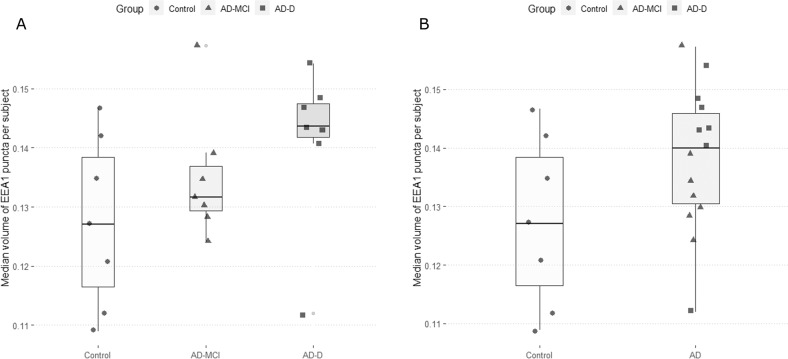
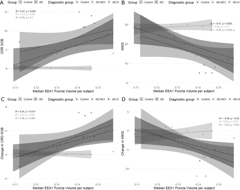
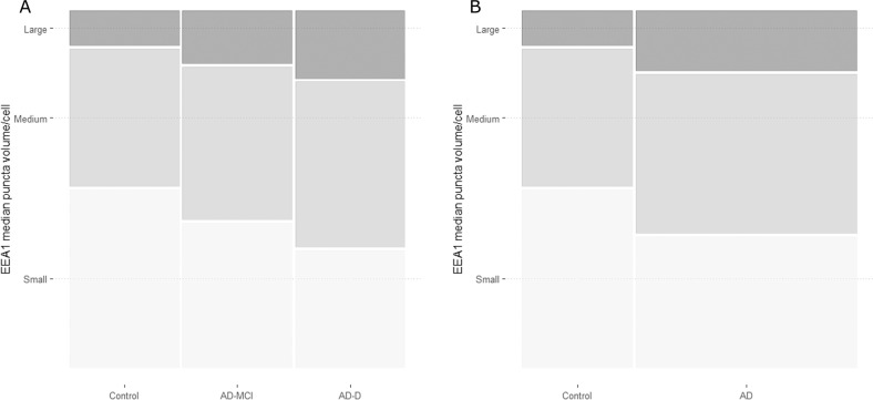
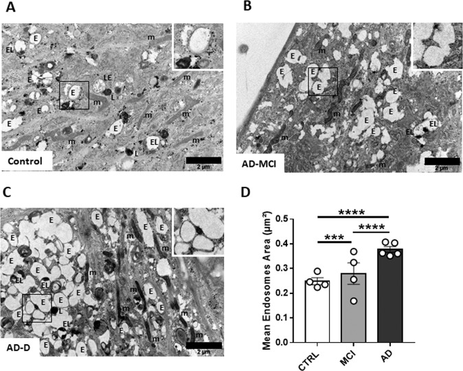
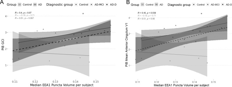
Morphological alterations of the endosomal compartment have been widely described in post-mortem brains from Alzheimer's disease (AD) patients and subjects with Down syndrome (DS) who are at high risk for AD. Immunostaining with antibodies against endosomal markers such as Early Endosome Antigen 1 (EEA1) revealed increased size of EEA1-positive puncta. In DS, peripheral cells such as peripheral blood mononuclear cells (PBMCs) and fibroblasts, share similar phenotype even in the absence of AD. We previously found that PBMCs from AD patients have larger EEA1-positive puncta, correlating with brain amyloid load. Here we analysed the endosomal compartment of fibroblasts from a very well characterised cohort of AD patients (IMABio3) who underwent thorough clinical, imaging and biomarkers assessments. Twenty-one subjects were included (7 AD with mild cognitive impairment (AD-MCI), 7 AD with dementia (AD-D) and 7 controls) who had amyloid-PET at baseline (PiB) and neuropsychological tests at baseline and close to skin biopsy. Fibroblasts isolated from skin biopsies were immunostained with anti-EEA1 antibody and imaged using a spinning disk microscope. Endosomal compartment ultrastructure was also analysed by electron microscopy. All fibroblast lines were genotyped and their AD risk factors identified. Our results show a trend to an increased EEA1-positive puncta volume in fibroblasts from AD-D as compared to controls (p.adj = 0.12) and reveal enhanced endosome area in fibroblasts from AD-MCI and AD-AD versus controls. Larger puncta size correlated with PiB retention in different brain areas and with worse cognitive scores at the time of biopsy as well as faster decline from baseline to the time of biopsy. Finally, we identified three genetic risk factors for AD (ABCA1, COX7C and MYO15A) that were associated with larger EEA1 puncta volume. In conclusion, the endosomal compartment in fibroblasts could be used as cellular peripheral biomarker for both amyloid deposition and cognitive decline in AD patients.
内体区室的形态改变已在阿尔茨海默病(AD)患者和唐氏综合征(DS)患者的死后大脑中广泛描述,这些患者是 AD 的高危人群。用针对内体标志物的抗体(如早期内体抗原 1(EEA1))进行免疫染色显示,EEA1 阳性斑点的大小增加。在 DS 中,外周细胞(如外周血单核细胞(PBMC)和成纤维细胞)即使没有 AD,也具有相似的表型。我们之前发现,AD 患者的 PBMC 具有更大的 EEA1 阳性斑点,与脑淀粉样蛋白负荷相关。在这里,我们分析了来自经过充分临床、影像和生物标志物评估的 AD 患者(IMABio3)的非常特征明确的队列的成纤维细胞的内体区室。纳入了 21 名受试者(7 名 AD 伴轻度认知障碍(AD-MCI),7 名 AD 伴痴呆(AD-D)和 7 名对照),他们在基线时进行了淀粉样蛋白-PET(PiB)和神经心理学测试,接近皮肤活检。用抗 EEA1 抗体对从皮肤活检中分离的成纤维细胞进行免疫染色,并使用旋转盘显微镜进行成像。还通过电子显微镜分析了内体区室的超微结构。对所有成纤维细胞系进行基因分型,并确定其 AD 危险因素。我们的结果显示,与对照组相比,AD-D 患者的成纤维细胞中 EEA1 阳性斑点的体积有增加的趋势(p.adj = 0.12),并且 AD-MCI 和 AD-AD 患者的成纤维细胞中的内体面积增大。较大的斑点大小与不同脑区的 PiB 保留以及活检时的认知评分更差以及从基线到活检时的更快下降相关。最后,我们确定了三个与 AD 相关的遗传危险因素(ABCA1、COX7C 和 MYO15A),这些危险因素与更大的 EEA1 斑点体积相关。总之,成纤维细胞中的内体区室可用作 AD 患者淀粉样蛋白沉积和认知能力下降的细胞外周生物标志物。